


A single C. elegans PUF protein binds RNA in multiple modes
- Yvonne Yiling Koh1,2,
- Laura Opperman1,4,
- Craig Stumpf1,
- Arpita Mandan1,5,
- Sunduz Keles3 and
- Marvin Wickens1
- 1Department of Biochemistry, University of Wisconsin–Madison, Madison, Wisconsin 53706, USA
- 2Microbiology Doctoral Training Program, University of Wisconsin–Madison, Madison, Wisconsin 53706, USA
- 3Departments of Statistics and Biostatistics and Medical Informatics, University of Wisconsin–Madison, Madison, Wisconsin 53706, USA
Abstract
PUF proteins specifically bind mRNAs to regulate their stability and translation. Here we focus on the RNA-binding specificity of a C. elegans PUF protein, PUF-11. Our findings reveal that PUF-11 binds RNA in multiple modes, in which the protein can accommodate variable spacings between two distinct recognition elements. We propose a structural model in which flexibility in the central region of the protein enables the protein to adopt at least two distinct structures, one of which results in base flipping.
Keywords
INTRODUCTION
RNA–protein interactions control every step in an mRNA's life. The specificities of regulatory proteins for RNA determine which mRNAs are controlled and how. The 3′ untranslated region (3′UTR) is a key repository for elements that govern stability, translation, and localization (Wickens et al. 2002). Several families of RNA-binding proteins have been identified that interact with such elements. Here we focus on the PUF family of RNA-binding proteins, chosen because of their biological importance and the striking modularity of their interactions with RNA.
PUF proteins are highly conserved RNA-binding proteins that bind to specific RNA sequences. Members of the PUF family include Drosophila melanogaster Pumilio and Caenorhabditis elegans fem-3 binding factor (FBF) (Zamore et al. 1997; Zhang et al. 1997). PUF proteins elicit repression or decay of the mRNAs to which they bind (Ahringer and Kimble 1991; Wreden et al. 1997; Wharton et al. 1998; Olivas and Parker 2000; Chagnovich and Lehmann 2001; Goldstrohm et al. 2006, 2007) (for review, see Wickens et al. 2002; Goldstrohm and Wickens 2008). Each PUF appears to interact with multiple mRNAs, from 40 to 220 in yeast (Gerber et al. 2004) to more than 1000 in humans (Galgano et al. 2008; Morris et al. 2008). Multiple natural targets of C. elegans PUF proteins have been identified by genetics, interaction tests, immunocytochemistry, and association in extracts (Zhang et al. 1997; Crittenden et al. 2002; Lamont et al. 2004; Thompson et al. 2005; Lee et al. 2006, 2007) (for review, see Kimble and Crittenden 2007). While the biological roles are diverse, the maintenance of diverse stem cells appears to be a common function (Wickens et al. 2002; Morrison and Kimble 2006; Kimble and Crittenden 2007), as may be its roles in the nervous system (Schweers et al. 2002; Dubnau et al. 2003; Ye et al. 2004; Chen et al. 2008).
PUF proteins have a reiterated repeat structure. Virtually all PUF proteins possess eight repeats, each of which comprises a three-helix domain of ∼40 amino acids (Zamore et al. 1997; Edwards et al. 2001; Wang et al. 2001). These domains lie on one another to form a ladder of eight α-helices, termed RNA recognition helices, which bind RNA. In the crystal structure of human Pumilio1 bound to RNA (Wang et al. 2002), specific contacts are formed between the eight consecutive recognition helices and nucleotides, yielding a one-helix/one-base binding mode (Fig. 1A). Each recognition helix uses several amino acid side chains to contact the cognate base via hydrogen bonds, van der Waals forces, and stacking interactions (Wang et al. 2002).
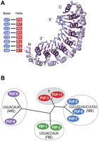
PUF proteins bind distinct sites. (A) Hs PUM1; structure of human Pumilio1–RNA complex (PDB accession code 1M8Y) (Wang et al. 2002). Eight RNA-recognition helices (purple in structure, red in diagram) contribute side chains to contact the RNA bases (blue). Hydrogen-bonding interactions are indicated by red lines in the structure; stacking interactions are not shown. (B) Four clusters of C. elegans PUF proteins (Stumpf et al. 2008b). PUF proteins are grouped based on amino acid similarity; known binding sites are indicated for each cluster.
The residues that form base-specific contacts in human Pumilio1 are highly conserved among all PUF proteins, yet not all PUF proteins bind the same sites (Gerber et al. 2004; Bernstein et al. 2005; Opperman et al. 2005; Seay et al. 2006; Stumpf et al. 2008b). The binding sites of C. elegans PUF-8 and FBF comprise a UGU and more 3′ AU element (Opperman et al. 2005). In the PUF-8 binding site, these two elements are spaced by 2 nucleotides (nt), and in FBF, by three. This distinction enables the two PUF proteins to efficiently bind only their own sites, yet use nearly identical sets of atomic contacts. The structure of FBF bound to RNA indicates that the additional base is turned away from the protein, or “flipped” (Y. Wang, L. Opperman, M. Wickens, and T. Hall, in prep.). Flipped bases appear to be a common principle used to achieve PUF specificity, as they are seen in yeast Puf4p complexes with natural target RNAs and in human Pumilio1 bound to noncognate RNA (Gupta et al. 2008; Miller et al. 2008). In both FBF and yeast Puf4p, the identity of the additional base is unimportant for protein binding (Miller et al. 2008; Y. Wang, L. Opperman, M. Wickens, and T. Hall, in prep.). Indeed, flexibility in nucleotide identity at a particular position in a PUF recognition site may be good prima facie evidence of flipping.
C. elegans possess 10 PUF proteins, which cluster into four groups based on sequence similarity: these are termed the FBF, PUF-8/-9, PUF-5/-6/-7, and PUF-3/-11 groups (Fig. 1B; Stumpf et al. 2008b). Each of the three groups analyzed thus far has a distinctive RNA-binding specificity, as indicated in Figure 1; each recognizes a UGU sequence at the 5′ end of the core site, as is typical of all validated PUF protein targets. Importantly, the biological functions of the different groups of worm PUF proteins are distinct, but overlap (e.g., Zhang et al. 1997; Lamont et al. 2004; Bachorik and Kimble 2005; Lublin and Evans 2007; Nolde et al. 2007). These findings imply that the different groups of proteins control distinct sets of mRNAs. Similarly, yeast Puf3p binds 220 mRNAs, yet few of these bind the other four canonical PUF proteins of that organism (Gerber et al. 2004).
In this study, we sought to determine the RNA-binding specificity of C. elegans PUF-11. Our data reveal surprising flexibility in PUF-11's sequence specificity, and suggest that this protein binds to RNA in multiple modes. We suggest that Puf-11–RNA complexes exist in at least two different conformations, which enable recognition of different groups of mRNAs. We propose a structural model for the interaction.
RESULTS
PUF-11 binds three classes of RNA sequences
To characterize the RNA-binding specificity of PUF-11, we performed a selection experiment using the yeast three-hybrid system (Fig. 2A; Hook et al. 2005; Stumpf et al. 2008a). We constructed a library of plasmids that encoded hybrid RNAs. The RNAs possessed a UGU trinucleotide, flanked by three randomized nucleotides upstream (5′) and seven randomized nucleotides downstream (3′). The library was introduced into yeast together with a plasmid encoding a PUF-11/AD fusion protein. RNAs that bound PUF-11 activated expression of the reporter gene, HIS3, and so were identified by growth on selective media (Fig. 2B). Secondary screens confirmed that the RNAs obtained also activated a second reporter gene, LacZ, and that they required the PUF-11/AD protein to do so (data not shown). A total of 66 unique RNA sequences were obtained (Fig. 2B).
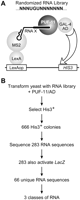
Identification of PUF-11 binding sites. (A) Yeast three-hybrid selection used to identify PUF-11 binding sites. The randomized region of the RNA library is illustrated using N to indicate a randomized base. Interaction between PUF-11 and RNA X will trigger expression of reporter genes HIS3 and LacZ. (B) Summary of three-hybrid selection experiment. A total of 666 positive clones were obtained. Of these, 283 clones were recovered from Escherichia coli and sequenced. Sixty-six unique sequences were present among the 283 clones, and these unique sequences yielded three classes of RNAs.
The set of sequences fit three position weight matrices (PWMs), and so yielded three classes of RNA sequences (Fig. 3; see Materials and Methods). Class I comprised 27 sequences, Class II, 25 sequences, and Class III, 16 sequences. We determined the relative binding affinities of all the RNAs to PUF-11 by assaying for β-galactosidase activity using the three-hybrid system. The magnitude of β-galactosidase activity in the system correlates with the affinity of the protein–RNA interaction (Hook et al. 2005). The range of β-galactosidase activities was similar in all three classes (Fig. 3). In particular, the high-affinity sites of each class bound with similar affinities.
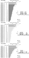
PUF-11 binds to three classes of RNAs. β-galactosidase activity as determined using strains possessing each of the indicated RNAs. β-galactosidase activity reflects the strength of PUF–RNA interactions (Hook et al. 2005). Values are relative to RNA I-1. The sequence in the relevant region of each bound RNA is indicated and corresponds to the portion that had been randomized. RNAs are named as X-Y, where X indicates the class, and Y indicates relative affinity for PUF-11 within the class. RNAs I-1, II-1, and III-1 were included when testing each class of RNAs, to provide standards. The consensus sequence for each class was derived using WebLogo software and displays information content and frequency of nucleotide occurrence at each position.
The three RNA classes differ in sequence. Each class possessed an AU dinucleotide downstream of the UGU trinucleotide (Fig. 3). The following properties were diagnostic of each class.
-
Class I: The AU dinucleotide was located in positions +6 and +7. An adenosine was highly preferred in position +5.
-
Class II: The AU dinucleotide was located in positions +7 and +8. The identities of bases between the UGU and AU elements were more degenerate than in Classes I and III. Class II RNAs were enriched for C at the -1 position; Classes I and III were not.
-
Class III: The AU dinucleotide was located in positions +7 and +8. An adenosine was highly preferred in position +5, as in Class I.
Taken together, these data suggest distinct modes of PUF-11 binding, which differ in the spacing of the UGU and AU elements, and in preferences at the −1 and +5 positions.
To compare affinities for the three classes quantitatively, we analyzed PUF-11 binding in vitro. We selected one RNA from each class that closely matched the consensus and had bound PUF-11 with high affinity in the three-hybrid assay. These three RNAs were used in an electrophoretic mobility-shift assay together with purified recombinant PUF-11 fused to glutathione S-transferase (GST) (Fig. 4A). GST–PUF-11 bound RNA I-1 with an apparent Kd of 43 ± 3 nM; RNA II-1, 62 ± 4 nM; and RNA III-1, 54 ± 5 nM (Fig. 4B). We conclude that the avidities of these RNAs for GST–PUF-11 were similar, corroborating the three-hybrid data.
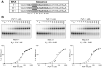
PUF-11 binds high-affinity sites of each RNA class with comparable affinities in vitro. (A) RNAs from each class that bound PUF-11 with high affinity in the three-hybrid assay. The sequences of the synthetic RNA oligonucleotides used for EMSAs are shown. The core binding element of each RNA is highlighted. Numbering begins with +1 at the first U of the core UGU trinucleotide. (B) EMSAs with purified recombinant GST–PUF-11. Protein concentrations are indicated above the gel. Two complexes of different electrophoretic mobilities were observed: The complex migrates slower than free RNA. The fraction of RNA bound was plotted against protein concentration to obtain apparent Kd values for each RNA.
Different specificities
To compare the RNA-binding specificity of PUF-11 to that of other C. elegans PUF proteins, we tested PUF-11 binding to the known binding sites of other PUF proteins, and the binding of other PUF proteins to PUF-11 binding sites. For this purpose, we used two RNAs from each class that bound with high and low affinities. Using the three-hybrid assay, we determined that PUF-11 bound the two sequences from each class as expected (Fig. 5). PUF-11 did not bind the PUF-8 site (NRE) and bound the PUF-5 site (5BE) only weakly. (Weak binding to the 5BE was due to an adventitious site in the vector sequence.) Conversely, PUF-5 bound its cognate site (the 5BE), but none of the PUF-11 sites (Fig. 5). PUF-8 bound its cognate site, the NRE; it also bound the Class I RNA, I-26. The interaction of PUF-8 with this RNA was expected, as the RNA possessed two key determinants of PUF-8 binding: an AU dinucleotide in positions +6 and +7 and an A at +4 (Opperman et al. 2005). Conversely, PUF-11 did not bind the NRE because the NRE has a C rather than an A at +5. We conclude that PUF-11's specificity is distinct from those of PUF-5 and PUF-8.
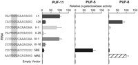
PUF proteins with different specificities. Three-hybrid analysis of different C. elegans PUF proteins. Relative binding of RNAs to PUF-11 was normalized to RNA I-1; binding to PUF-5 was normalized to 5BE; and binding to PUF-8 was normalized to NRE.
Different modes of RNA binding
Class I and Class III RNAs generally possessed an A at position +5. To test the role of this position in greater depth, we analyzed base substitutions at this position in each representative RNA using the three-hybrid assay (Fig. 6A). In RNA I-1, an adenosine was absolutely required for binding: any substitution reduced binding to background levels. However, in RNA II-1, no such requirement was observed: any base was tolerable. RNA III-1 was intermediate: either purine supported binding, while both pyrimidines did not. To quantify the effects of the +5 base identity, we analyzed binding of GST–PUF-11 in vitro to RNAs of each class that possessed either A or C at this position (Fig. 6B). The data confirm the three-hybrid results: In RNA I-1 and RNA III-1, a +5A was strongly preferred, but in RNA II-1, A or C bound equally well. We infer that PUF-11 may recognize the base at position +5 of Classes I and III, but not Class II, again suggesting different modes of RNA binding.
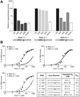
Mutational analysis of position +5 in PUF-11 binding sites. (A) Base substitutions at position +5 of three RNAs analyzed using the three-hybrid assay. The core binding element of each RNA is shown; UGU is shaded and the mutated base is underlined. β-galactosidase values are relative to the respective wild-type RNAs. (B) Base substitutions at position +5 of three RNAs analyzed in vitro. Synthetic RNAs with the indicated base substitution at position +5 were analyzed in EMSAs with purified recombinant GST–PUF-11. Krel is the ratio of the Kd relative to the respective wild-type RNAs.
Class II RNAs generally possessed a C at position −1. To test the role of this nucleotide systematically, we analyzed base substitutions at this position in each representative RNA (Fig. 7). In both three-hybrid (Fig. 7A) and gel-shift assays (Fig. 7B), a C at position −1 was important for PUF-11 binding to RNA I-1 and RNA II-1, but not for binding to RNA III-1. In I-1 and II-1, both pyrimidines permitted binding, but C was preferred (Fig. 7A). The preference for a −1C in Class I RNAs was surprising, since that preference was not observed in the three-hybrid selection experiments (Fig. 2); this likely reflects the stringency of the selection, which permitted detection of weaker Class I binding sites. Taken together, our results suggest that PUF-11 recognizes the base at the −1 position in Class I and Class II RNAs, but not in Class III.
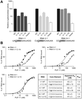
Mutational analysis of position -1 in PUF-11 binding sites. (A) Base substitutions at position −1 of three RNAs analyzed using the three-hybrid assay. The core binding element of each RNA is shown; UGU is shaded and the mutated base is underlined. β-galactosidase values are relative to the respective wild-type RNAs. (B) Base substitutions at position −1 of three RNAs analyzed in vitro. Synthetic RNAs with the indicated base substitution at position −1 were analyzed in EMSAs with purified recombinant GST–PUF-11. Krel is the ratio of the Kd relative to the respective wild-type RNAs.
Our results support the following conclusions: The identities of bases at −1 and +5 are important determinants of binding, and the requirements vary among classes of binding sites. These findings support the hypothesis that PUF-11 can bind RNA in multiple modes.
Alignment of RNA and protein
The presence of UGU and AU elements in all three RNA classes suggest that PUF-11 forms base-specific contacts with this element. To align specific PUF-11 repeats with the different classes of RNAs, we analyzed compensatory mutations between the protein and the RNAs (Figs. 8–10). The base specificity of PUF repeats can be switched rationally, by changing the identities of specific amino acid side chains that make edge-on contacts with the bases (Wang et al. 2002). This manipulation makes it possible to assign specific helices to the bases they recognize. In several PUF proteins, the three conserved residues of the RNA-recognition helix in Repeat 7 specify a guanosine (Wang et al. 2002; Miller et al. 2008; Y. Wang, L. Opperman, M. Wickens, and T. Hall, in prep.; D. Zhu, C. Stumpf, M. Wickens, and T. Hall, in prep.). To construct PUF-11 mutant proteins, we substituted these three residues (serine, histidine, and glutamate) at analogous positions in other repeats, and tested those mutant proteins against RNAs containing mutations at specific bases. In this fashion, we altered the base specificities of repeats 2, 3, and 4 in PUF-11, and aligned their RNA-recognition helices with different RNAs.
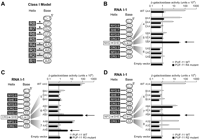
Alignment of PUF-11 repeats and RNA I-1. (A) A model illustrating how each PUF-11 repeat (R1–R8, shaded) could interact with eight bases (A8–U1, unshaded) in the core binding element of RNA I-1. Black lines indicate hydrogen bonds; stacking interactions and van der Waals forces are not shown. Triangles indicate conserved interactions among PUF proteins; asterisks and a gray box indicate mapped interactions between PUF-11 and RNA I-1 based on B–D. (B–D) Binding of Repeats 4, 3, and 2 mutant proteins to RNA I-1. Binding scheme is shown on the left, with compensatory protein and RNA mutations unshaded. Binding of wild-type and mutant proteins to RNAs using the three-hybrid assay is shown on the right. The new interactions that map the base to the α-helix are indicated by arrows.
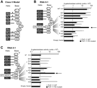
Alignment of PUF-11 repeats and RNA II-1. (A) A model illustrating how each PUF-11 repeat (R1–R8, shaded) could interact with nine bases (A9–U1, unshaded) in the core binding element of RNA II-1. Black lines indicate hydrogen bonds; stacking interactions and van der Waals forces are not shown. Triangles indicate conserved interactions among PUF proteins; asterisks and a gray box indicate mapped interactions between PUF-11 and RNA II-1 based on B and C. (B,C) Binding of Repeats 3 and 2 mutant proteins to RNA II-1. Binding scheme shown on left, with compensatory protein and RNA mutations unshaded. Binding of wild-type and mutant proteins to RNAs using the three-hybrid assay is shown on the right. The new interactions that map the base to the α-helix are indicated by arrows.
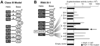
Alignment of PUF-11 repeats and RNA III-1. (A) A model illustrating how each PUF-11 repeat (R1–R8, shaded) could interact with nine bases (A9–U1, unshaded) in the core binding element of RNA III-1. Black lines indicate hydrogen bonds; stacking interactions and van der Waals forces are not shown. Triangles indicate conserved interactions among PUF proteins; asterisks and a gray box indicate mapped interactions between PUF-11 and RNA III-1 based on B. (B) Binding of Repeat 2 mutant protein to RNA III-1. Binding scheme is shown on the left, with compensatory protein and RNA mutations unshaded. Binding of wild-type and mutant proteins to RNAs using the three-hybrid assay is shown on the right. The new interactions that map the base to the α-helix are indicated by arrows.
Class I RNAs (Fig. 8)
Class I RNAs possess an AU in positions 6 and 7, as do RNAs bound by human Pumilio1 (Fig. 8A). Thus, we predicted that PUF-11 bound RNA in an uninterrupted one-helix/one-base fashion, as does Pumilio (Fig. 8A). To align Repeat 4 with RNA I-1, we mutated its Asn-His-Gln to Ser-His-Glu, and then tested RNA base specificity at positions 4, 5, or 6 of RNA I-1 (Fig. 8B). If PUF-11 does bind in the same manner as human Pumilio1, then the Repeat 4 mutant protein should bind an RNA with a G at nucleotide 5 (rather than an A, as in wild type). We tested the mutant protein with wild-type RNA I-1 and nine other RNAs, each containing a single-base substitution at positions 4, 5, or 6 (Fig. 8B). Wild-type PUF-11 bound RNAs with an A, U, or G at position 4, and required A at both positions 5 and 6. The Repeat 4 mutant protein, however, required a G at nucleotide 5 and did not bind wild-type or other mutant RNAs. Thus, switching Asn-His-Gln to Ser-His-Glu changed the specificity from A to G at nucleotide 5. Based on these data, we conclude that Repeat 4 of PUF-11 recognizes nucleotide 5 of RNA I-1.
We used the same strategy to align Repeat 3 with RNA I-1 (Fig. 8C). We predicted that switching Cys-Arg-Gln to Ser-His-Glu would change the specificity for nucleotide 6 from A to G. Indeed, the Repeat 3 mutant protein bound when nucleotide 6 was a G, which the wild-type protein did not bind. Both proteins still accepted A at that position. Thus, we positioned Repeat 3 opposite of nucleotide 6 in RNA I-1. Surprisingly, the mutant protein bound the RNA when nucleotide 7 was a G, which the wild-type protein did not. Either Repeat 3 toggles between nucleotides 6 and 7 or the mutation in the stacking amino acid also changes identity in the adjacent base. Indeed, arginine stacks poorly on guanosine, and that may be relieved by the His substitution (Morozova et al. 2006).
To align repeat 2 with RNA I-1, we changed Asn-Phe-Gln to Ser-His-Glu (Fig. 8D). The repeat 2 mutant protein bound when nucleotide 7 was a G, which the wild-type protein did not. Both proteins still accepted A at that position. Thus, we were able to position repeat 2 opposite of nucleotide 7 in RNA I-1.
Based on these compensatory mutations, we positioned repeats 2, 3, and 4 opposite nucleotides 7, 6, and 5, respectively (Fig. 8A). We conclude that PUF-11 bound RNA I-1 in a continuous one-helix/one-base manner as does human Pumilio1.
Class II RNAs (Fig. 9)
In Class II RNAs, the AU dinucleotide lies at positions 7 and 8, 1 nt further from the UGU than in Class I. Nonetheless, we hypothesized that Repeats 2 and 3 might still recognize the AU element (Fig. 9A). To align Repeat 3 with RNA II-1, we changed Cys-Arg-Gln to Ser-His-Glu (Fig. 9B) and predicted that it would now bind G rather than A. Indeed, that was the case: The Repeat 3 mutant protein preferred G (and accepted A), while the wild type-protein preferred A. Thus, we positioned repeat 3 opposite nucleotide 7 in RNA II-1.
To align Repeat 2 with RNA II-1, we changed Asn-Phe-Gln to Ser-His-Glu (Fig. 9C). We predicted that the specificity for nucleotide 8 would change from U to G. Wild-type protein preferred U over the other bases at nucleotide 8, but the Repeat 2 mutant protein preferred G (and A). We conclude that Repeat 2 binds nucleotide 8 in RNA II-1. Together, these results indicate that PUF-11 accommodates an additional base in RNA II-1 as compared with RNA I-1. This mode of binding was distinct from that in RNA I-1, where eight repeats contact 8 nt.
Class III RNAs (Fig. 10)
The spacing in Class III RNAs is the same as in Class II (Fig. 10A). To align Repeat 2 with RNA III-1, we changed Asn-Phe-Gln to Ser-His-Glu (Fig. 10B). We predicted the specificity for nucleotide 8 would change from U to G, and, indeed, this was the case. Thus, Repeat 2 lies opposite nucleotide 8 in RNA III-1.
Summary
The compensatory mutant strategy enabled us to align repeats with RNA in all three classes. Invariably, PUF-11 Repeats 2 and 3 recognized the AU dinucleotide, but at different nucleotide locations relative to UGU. PUF-11 bound RNA I-1 in an uninterrupted one-helix/one-base manner, but accommodated an additional nucleotide in RNA II-1 and RNA III-1. We conclude that PUF-11 employs distinct RNA-binding modes with different RNAs.
DISCUSSION
PUF-11 binds to RNA in at least two modes, defined by differences in the spacing of two recognition elements, UGU and AU, and preferences for nucleotides at −1 and +5. A cytosine at −1 and an adenosine at +5 are preferred in certain contexts. The distance between the UGU and AU elements can either be 2 or 3 nt; the difference in spacing has little effect on affinity. Compensatory mutants reveal that the same helices recognize AU, and presumably the UGU, in each mode.
We propose a structural model of PUF-11 interactions with RNA. This model focuses on flexibility in the central region of the protein and base flipping (Fig. 11), and draws on the known structures of human Pumilio1 and C. elegans FBF. In the “curved and in” conformation, the central region of PUF-11 is curved as in human Pumilio1, resulting in a one-to-one alignment of bases and α-helices, and no base flipping (Fig. 11, left). In the “flat and flipped” conformation, PUF-11 is flattened in the region opposite nucleotides +5 and +6, resulting in base flipping (Fig. 11, right). These two states are analogous to those seen in complexes of human Pumilio1 with cognate RNA, which is “curved and in” (Wang et al. 2002), and of FBF-2, which is “flat and flipped” (Y. Wang, L. Opperman, M. Wickens, and T. Hall, in prep.). Similarly, human Pumilio1 can bind noncognate RNA in a manner that involves base flipping (Gupta et al. 2008). It is striking that PUF-11 binds with comparable affinity in either mode, implying that base-flipping does not result in a dramatic energy cost, despite the presumed loss of specific contacts with the protein. Base-to-base stacking likely compensates.
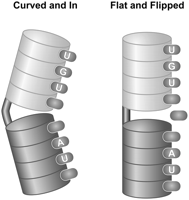
Model of PUF-11 interactions with RNA. The protein is depicted as a series of discs corresponding to PUF repeats, arranged in two groups of four. Interactions of bases (ellipse) with binding pockets in each repeat also are shown. Alternate conformations in the central region of the protein alter the overall curvature, as has been observed with FBF (Y. Wang, L. Opperman, M. Wickens, and T. Hall, in prep.), and affect base flipping. In one conformation, the protein is curved and the bases are “in,” contacting the protein (left, “Curved and In”). In the other conformation, the protein is flat and the central bases flip (right, “Flat and Flipped”). The alteration of structure may involve different helical packing in that region, rather than a distinct “hinge” region. See the text for details.
In FBF and yeast Puf4p, the identities of flipped bases are flexible, while the identities of bases that make protein-specific contacts are constrained (Miller et al. 2008; Y. Wang, L. Opperman, M. Wickens, and T. Hall, in prep.). In Class I RNAs, the highly conserved bases between UGU and AU suggest that these nucleotides form specific contacts with PUF-11 in a continuous one-helix/one-base manner (as in human Pumilio1). In Classes II and III, however, the degeneracy of bases between the two motifs suggests that one or more nucleotides turn away from the protein. UGU invariably is recognized by Repeats 6–8 of PUF-11, and AU by Repeats 2 and 3.
In one simple view, interaction with one element, say the UGU, would enable a “search” for additional recognition elements nearby. Upon that second interaction, with the AU dinucleotide, bases that do not interact with the protein are left stacked on one another. It is unclear whether these two protein conformations exist in the apo-protein, or are induced upon binding the RNA. In either case, the flattened protein may be able to accommodate an even greater number of additional bases between the UGU and AU.
In Classes II and III RNAs, UGU and AU are spaced identically; the distinction between these classes may lie in precisely which central bases contact the protein. In Class II RNAs, the tolerance of PUF-11 for all bases at position +5 indicates that base may flip; in Class III RNAs, the preference for A at position +5 suggests that it contacts PUF-11, which implies that +4 may flip. In both instances, the contact is likely to be with the same binding pocket of Repeat 5. Indeed, this is likely to be the same site to which the adenosine (or guanosine) at +4 binds in Class I RNAs.
The distinctive features of complexes with Class I, II, and III RNAs may enable them to be used differentially in vivo. For example, proteins that interact with the PUF-RNA complexes in vivo, such as CPEB-1, NANOS-3, and GLD-2/GLD-3, could readily discriminate one class from another. Protein partners might also restrict flexibility of the apo-protein, driving its interaction with specific mRNAs.
The −1 position presents another key facet of specificity, in which PUF-11 strongly prefers a cytosine in Classes I and II RNAs. In this respect, PUF-11 echoes yeast Puf3p; a binding pocket at the C terminus of the Puf3p RNA-binding domain specifically recognizes a C at the −2 position (D. Zhu, C. Stumpf, M. Wickens, and T. Hall, in prep.). We suggest that PUF-11 possesses an analogous binding pocket for a cytosine. The identity of the −1 position was unimportant in binding to Class III RNAs; this may be due to the use of the cytosines at either −2 or −3 in the place of −1. Alternatively, the precise interactions in positions +4 to +6 could affect the pocket that recognizes the −1C.
Our data demonstrate that a single PUF protein recognizes different sites using distinct modes of RNA binding. This greatly diversifies the possibilities for regulation, and the potential effects of coregulators. Structural analysis will reveal the precise nature of the complexes on the three classes of RNA and will open the door to mechanistic studies of how those different complexes form and are used for regulation in vivo.
MATERIALS AND METHODS
Constructs for yeast three-hybrid assay
The RNA-binding region of PUF-11 (GenBank accession no. NM_171363), comprising amino acids 50–505, was cloned into an activation domain vector, pGADT7 (Stumpf et al. 2008b). For specificity testing of different PUFs (Fig. 5), full-length PUF-5 (NM_063413) and residues 127–519 of PUF-8 (NM_063122) were cloned into pACTII activation vectors. The PUF-11 mutants used in the compensatory experiments (Figs. 8–10) were created by site-directed mutagenesis. Amino acid changes were as follows: for PUF-11 Repeat 4 mutant, N272S and Q276E; Repeat 3 mutant, C235S, R236H, and Q239E; Repeat 2 mutant, N199S, F200H, and Q203E. The randomized RNA library was constructed by cloning DNA oligonucleotides (CCGGCTAGCNNNTGTNNNNNNNAATTTAATAAAGCATG) into the XmaI and SphI sites of p3HR2 (Stumpf et al. 2008b). All other RNA constructs used in the three-hybrid assay were created in the same fashion.
Yeast three-hybrid assay
Three-hybrid assays were performed in the YBZ-1 yeast strain as described previously (Hook et al. 2005). Selection experiments were carried out using medium lacking histidine and in the presence of 5 mM 3-aminotriazole. A total of 283 out of 666 positive interactors were recovered in Escherichia coli and reintroduced into naïve YBZ-1 to confirm the interaction and specificity. Three-hybrid assays were conducted with the following modifications: 100 μL of a saturated culture was diluted into 4-mL selective medium and allowed to grow for 2.5 h to reach an OD660 of 0.1–0.2. β-galactosidase activity was normalized to an OD660 of 0.1. β-galactosidase values are represented as an average of three repetitions, with standard deviation shown in each figure.
Protein construct and purification
PUF-11 (amino acids 50–505) was cloned into pGEX6P1 (Amersham) as GST fusion proteins and purified as described previously (Hook et al. 2005). Protein was eluted from the glutathione-sepharose resin (Amersham) without cleavage and dialysis. Elution buffer consists of 50 mM reduced glutathione (Sigma), 1X PBS, 0.2% Tween-20, 150 mM NaCl, 0.1% β-mercaptoethanol, and 30% glycerol (pH 8.0). Protein purity was measured by SDS gel electrophoresis and concentration determined by a Bradford assay.
Electrophoretic mobility-shift assay
A total of 100 fmol of RNA oligonucleotides (Dharmacon), 32P 5′ end-labeled using T4 kinase, were combined with a range of protein concentrations in 10 mM HEPES (pH 7.4), 1 mM EDTA, 20 mM NaCl, 1 mM DTT, and 0.002% Tween-20 for 30 min at room temperature. Loading dye (5 μL of 6% glycerol and 0.06% Bromophenol Blue) was added to each 10-μL reaction before loading 5 μL on a pre-run nondenaturing 5% polyacrylamide gel (BioRad). Gels were resolved in 1X TBE at 100 V for 30 min at 4°C. Gels were then exposed to storage phosphor screens for 1 h and scanned using a Typhoon 9410 Workstation (GE Healthcare). The fraction of bound RNA relative to total RNA in the reaction was determined using ImageQuant (Amersham). The apparent Kd, concentration of protein at which half-maximal binding occurs, was determined using GraphPad Prism 4. The Kd values and standard errors reported here were from four independent experiments.
Computational analysis of sequences
We used a mixture of position weight matrices (PWMs) (Barash et al. 2003) to characterize whether these sets of sequences are best described by multiple RNA sequence classes. This probabilistic model assumed that each sequence originated from one of the K RNA sequence classes and model fitting involved inferring the class of each sequence as well as choosing the best K. We considered values between 1 and 6 for K and multiple model selection criteria yielded K = 3 as the optimal number.
ACKNOWLEDGMENTS
We appreciate the advice and comments of members of the Wickens laboratory and the help of Laura Vanderploeg in preparing the figures. Y.K. is supported by the A*STAR National Science Scholarship from Singapore, S.K. by NIH grant HG003747, and M.W. by NIH grants 144-MK47 and 144-QS25. A.M. was supported by a Khorana Summer Fellowship during her participation in these studies.
Footnotes
-
↵4 Present addresses: Department of Genetics, University of Wisconsin–Madison, Madison, WI 53706, USA
-
↵5 Department of Chemistry, Indian Institute of Technology, Kalyanpur, India 208016.
-
Reprint requests to: Marvin Wickens, Department of Biochemistry, University of Wisconsin–Madison, Madison, WI 53706, USA; e-mail: wickens{at}biochem.wisc.edu; fax: (608) 262-9108.
-
Article published online ahead of print. Article and publication date are at http://www.rnajournal.org/cgi/doi/10.1261/rna.1545309.
-
- Received January 7, 2009.
- Accepted March 2, 2009.
- Copyright © 2009 RNA Society




