


Probing the architecture of the B. subtilis RNase P holoenzyme active site by cross-linking and affinity cleavage
Abstract
Bacterial ribonuclease P (RNase P) is a ribonucleoprotein complex composed of one catalytic RNA (PRNA) and one protein subunit (P protein) that together catalyze the 5′ maturation of precursor tRNA. High-resolution X-ray crystal structures of the individual P protein and PRNA components from several species have been determined, and structural models of the RNase P holoenzyme have been proposed. However, holoenzyme models have been limited by a lack of distance constraints between P protein and PRNA in the holoenzyme–substrate complex. Here, we report the results of extensive cross-linking and affinity cleavage experiments using single-cysteine P protein variants derivatized with either azidophenacyl bromide or 5-iodoacetamido-1,10-o-phenanthroline to determine distance constraints and to model the Bacillus subtilis holoenzyme–substrate complex. These data indicate that the evolutionarily conserved RNR motif of P protein is located near (<15 Å) the pre-tRNA cleavage site, the base of the pre-tRNA acceptor stem and helix P4 of PRNA, the putative active site of the enzyme. In addition, the metal binding loop and N-terminal region of the P protein are proximal to the P3 stem–loop of PRNA. Studies using heterologous holoenzymes composed of covalently modified B. subtilis P protein and Escherichia coli M1 RNA indicate that P protein binds similarly to both RNAs. Together, these data indicate that P protein is positioned close to the RNase P active site and may play a role in organizing the RNase P active site.
Keywords
INTRODUCTION
Ribonucleoproteins (RNPs), a large and diverse group of macromolecular complexes composed of both RNA and protein components, are involved in numerous cellular processes (e.g., Greider and Blackburn 1987; Green and Noller 1997; Blackburn 1999; Ramakrishnan 2002; Doudna and Batey 2004; Meister and Tuschl 2004; Meier 2005). In light of the ubiquity and diversity of RNPs, a key challenge is to understand how functions arise from the synergistic interaction of RNA and protein components. In this context, the study of bacterial ribonuclease P (RNase P) occupies a unique niche. Throughout phylogeny, RNase P is an essential RNP endonuclease that catalyzes site-specific cleavage of precursor tRNA (pre-tRNA) molecules to remove 5′ leader sequences (Pace and Brown 1995; Kurz and Fierke 2000; Christian et al. 2002b; Xiao et al. 2002; Harris and Christian 2003; Hsieh et al. 2004). In Eukaryotes and some Archaea, RNase P contains one RNA and multiple protein subunits whose functions are unclear (Xiao et al. 2002). In contrast, Bacterial RNase P is composed of one RNA (PRNA, ∼400 nucleotides [nt]) and one small protein subunit (P protein, ∼120 amino acids). PRNA alone can recognize, bind, and cleave pre-tRNA in vitro in the presence of high salt concentrations (Kole et al. 1980; Guerrier-Takada et al. 1983; Reich et al. 1988); however, P protein significantly influences multiple aspects of catalysis, and is required for function in vivo (Hsieh et al. 2004, and references therein). Together with a significant body of biochemical characterization, these features make bacterial RNase P an intriguing yet relatively simple model in which to study the functional and structural synergy between the components of a ribonucleoprotein complex.
While the RNA component of RNase P can catalyze pre-tRNA cleavage, P protein plays a central role in RNase P function. P protein interacts with the 5′ leader of pre-tRNA substrates, leading to enhanced affinity for substrates (Crary et al. 1998; Kurz et al. 1998; Niranjanakumari et al. 1998b; Stams et al. 1998; Christian et al. 2002b). In addition, P protein increases metal ion affinity (Kurz and Fierke 2002), and pre-tRNA cleavage rate constants (Kurz et al. 1998; Sun et al. 2006). Interestingly, the specific influence observed for P protein is strongly dependent upon the identity of the pre-tRNA substrate examined, suggesting that leader/protein interactions may modulate substrate processing efficiency in vivo (Sun et al. 2006). In addition, P protein-specific binding enhancements are observed for substrates with leaders that are five or more nucleotides in length (Crary et al. 1998; Rueda et al. 2005). An interaction with the pre-tRNA at the N(−5) position, would require the RNase P active site to be within ∼20 Å of the RNA–protein interface.
X-ray crystal structures of P protein from Bacillus subtilis (Stams et al. 1998) and Thermatoga maritima (Kazantsev et al. 2003) and the NMR structure of P protein from Staphylococcus aureus (Spitzfaden et al. 2000) reveal a remarkably similar overall topology for P proteins from different species, despite low sequence similarity. Three regions of the protein have been proposed to interact with RNA (Fig. 1): the RNR motif, the central cleft, and the metal binding loop. The RNR motif is a highly basic region located in an unusual left-handed βαβ crossover connection from β-strand 3 to helix B to β-strand 4 (K52 to R68, B. subtilis numbering), and is highly conserved among all bacterial P proteins, suggesting that this region is functionally important (Altman 1989; Pace and Brown 1995; Gopalan et al. 1997; Brown 1998; Jovanovic et al. 2002). Second, a large central cleft (CC), formed by helix A and the face of the central β sheet, which is surrounded by positively charged residues, has been demonstrated to interact with pre-tRNA leader sequences through a variety of biochemical assays (Gopalan et al. 1997; Crary et al. 1998; Kurz et al. 1998; Niranjanakumari et al. 1998b; Stams et al. 1998; Kurz and Fierke 2002; Rueda et al. 2005). Third, a highly variable loop that binds two zinc ions in the B. subtilis P protein crystal (including Q38, E40, and D42) (Stams et al. 1998), termed the metal binding loop (MBL), may interact with RNA by hydrogen bonding or through a coordinated metal ion.
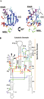
Structures of B. subtilis PRNA and B. subtilis P protein. (A) Structure of B. subtilis P protein indicating sites of single-cysteine mutations. The protein backbone is shown in blue with the RNR motif highlighted in magenta. Labels indicate the location of the RNR motif (RNR), metal binding loop (MBL), and central cleft (CC). Amino acids replaced with single-cysteine mutations near the N terminus (red), metal binding loop (green), RNR motif (black), β-strand 3 (yellow), and central cleft (cyan) are shown. (B) Secondary structure of the catalytic domain of B. subtilis PRNA, based on topology from the X-ray structure of the B. stearothermophilus PRNA (Kazantsev et al. 2005), indicating cleavage sites for AOP-labeled single-cysteine P protein variants: (arrow) RNR motif and β-strand 3 (red=R60, N61, K64; white=N61, N62; green=V46, N61, R65, R68; dark blue=R62; light blue=K64); (star) metal binding loop and β-strand 3 (black=Q38, E40, D42, R45; pink=Q38; purple=D42); and (diamond) N terminus (black=H3, K5, K7, K11, K12; white=H3, R7; orange=H3; aqua=K12) (summarized in Table 2). Shaded markers denote strong cleavage bands; open markers denote weaker cleavage bands. Highlighted nucleotides indicate sites where P protein reduced (red), enhances (blue), or has a variable effect (green) on cleavage during in-gel Fe(II)–EDTA cleavage assays (Buck et al. 2005b).
In light of its functional importance and potential proximity to the active site, a number of previous studies have investigated the positioning of the P protein within the RNase P holoenzyme. These studies have relied predominantly on Fe(II)–EDTA footprinting and chemical protection, and have been carried out in solution (Vioque et al. 1988; Chen et al. 1998; Massire et al. 1998; Biswas et al. 2000; Tsai et al. 2003), and with complexes separated by nondenaturing gel electrophoresis (Buck et al. 2005b). The orientation of the protein with respect to RNA has also been investigated using EPR and site-specific spin labeling (Gopalan et al. 1999). In addition, photocrosslinking has shown that the P protein central cleft is proximal to the substrate leader sequences (Niranjanakumari et al. 1998b). Together, these observations have allowed positioning of P protein in low-resolution models of the RNase P holoenzyme, and more recently in models based on crystal structures of PRNA in the absence of P protein and substrate (Buck et al. 2005b; Kazantsev et al. 2005; Torres-Larios et al. 2005). PRNA consists of two independently folding domains: the catalytic domain (C domain), containing the region required for catalytic activity, and the specificity domain (S domain), which contributes to pre-tRNA affinity (Fig. 1; Pace and Brown 1995; Pan 1995; Loria and Pan 1996). In general, previous data suggest that P protein contacts the catalytic domain of PRNA (Fig. 1), with specific contacts likely to involve the P1–P4 four-way helix junction in a region that has been proposed to contain the RNase P active site (Day-Storms et al. 2004; Buck et al. 2005b; Kazantsev et al. 2005; Torres-Larios et al. 2005).
While previous modeling studies have allowed for a general placement of P protein in structural models of RNase P, they are limited by the low (>20 Å) resolution and limited number of distance constraints provided by Fe(II)–EDTA cleavage, and the inability of chemical protection experiments to provide firm distance constraints between specific residues of P protein and PRNA. In addition, these previous studies were carried out in the absence of pre-tRNA substrates. To overcome these limitations and further explore the architecture of the RNase P holoenzyme–substrate complex, we have carried out extensive photocrosslinking and hydroxyl-radical cleavage of B. subtilis RNase P using covalently tethered reagents. These data identify regions of P protein proximal to PRNA and pre-tRNA and identify nucleotides near specific P protein amino acids. The resulting distance constraints were used to refine a biochemical model of the B. subtilis RNase P holoenzyme–substrate complex with respect to the positioning of P protein. Our data demonstrate that the P protein RNR motif, pre-tRNA cleavage site, and the putative active site elements of PRNA are located within a 15 Å radius.
RESULTS
Construction and characterization of single-cysteine P protein variants
To investigate the positioning of P protein in the RNase P holoenzyme and RNase P–pre-tRNAAsp complexes, we carried out photocrosslinking and hydroxyl-radical directed cleavage assays. For these studies, we constructed a library of 25 P protein variants with unique cysteine residues located throughout the protein, including the RNR motif, N terminus, and metal binding loop (Fig. 1). These single-cysteine variants provide unique sites for modification with either an azidophenacyl (AzP) group for photocrosslinking studies or acetamido-1,10-o-phenanthroline (AOP) for footprinting studies to identify nucleotides near P protein amino acids (Burgin and Pace 1990; Sigman and Chen 1990). Both of these reagents are proposed to react with RNA via direct contact (Burgin and Pace 1990; Sigman and Chen 1990) and thereby identify nucleotides within a 10–15 Å radius of the unique cysteine.
The success of structural studies using photoaffinity cross-linking and hydroxyl-radical cleavage strategies relies on the ability to derivatize P protein without significantly affecting holoenzyme structure. To assess the impact of single-cysteine mutations and subsequent derivatization on the formation of functional enzyme–substrate complexes, we measured the single turnover kinetics of RNase P containing mutated and AOP-modified proteins. We limited this analysis to mutants with cleavage patterns representative of their respective regions. For these experiments, RNase P was reconstituted with either a single-cysteine P protein mutant, or with AOP-modified single-cysteine mutants, and single turnover rate constants for cleavage of B. subtilis pre-tRNAAsp were determined at saturating enzyme concentrations (Table 1).
Single turnover cleavage rate constants for RNase P holoenzymes reconstituted with single-cysteine P protein variantsa
As shown in Table 1, single-cysteine mutations at representative locations in the N terminus (H3C, R7C, D15C), metal binding loop (Q38C, E40C, D42C), β-strand 3 (R45C, V46C), and RNR motif (R60C, N61C, R62C, K64C, R65C, R68C) have little or no effect on the single turnover cleavage rate constant, indicating the absence of structural alterations that affect cleavage efficiency. Similarly, cysteine mutation and AOP modification alters the cleavage rate constant less than twofold for all positions, except for R60C, which decreases the cleavage rate constant 10-fold (Table 1). Therefore, care must be taken in interpreting cross-linking and affinity cleavage results for position 60. It is notable in this regard that positions 60 and 65 are part of the conserved RNR motif (Gopalan et al. 1997; Jovanovic et al. 2002). The conservation and unusual topology of this region, together with observed rate defects for AOP-modified mutants in and near this region, are consistent with this region being located near the enzyme active site (see below).
Mapping the P protein–PRNA interface
Cross-linking of P protein variants to PRNA
Using the single-cysteine mutants described above, we identified regions of P protein near PRNA in the RNase P•pre-tRNAAsp complex using photocrosslinking. For these experiments, single-cysteine mutants were derivatized with azidophenacyl bromide (AzP). Phenylazide-mediated photocrosslinking requires close proximity of the photogenerated reactive species and its target; however, the cross-linked region can be as far away as 11 Å due to the length of the linker between the α-carbon of the modified cysteine and the reactive atom of the phenyl nitrene (Chen et al. 1993). For these experiments, the RNase P holoenzyme•substrate complex was reconstituted using stoichiometric amounts of AzP-labeled P protein and 5′-end-labeled PRNA in the presence of saturating concentrations of B. subtilis pre-tRNAAsp. In addition, the experiments were performed in the presence of CaCl2, which supports PRNA folding and substrate binding but decreases the hydrolytic rate constant 104-fold (Smith et al. 1992; Kurz et al. 1998). Complexes were irradiated with ultraviolet light (312 nm), and cross-linked species were separated by SDS-PAGE.
As shown in Figure 2, slow migrating bands indicative of intermolecular cross-links were observed for RNase P•pre-tRNAAsp complexes containing an AzP moiety attached to the N-terminal region (H3C, K5C, K6C, R7C, R9C, K12C, and D15C) and the metal binding loop (Q38C, E40C, and D42C) of P protein. In the RNR motif, AzP modifications at positions R60C, R65C, and R68C also result in cross-links to PRNA. No cross-link bands were observed in the absence of AzP modification or UV irradiation (data not shown; Fig. 2). In addition, no intermolecular cross-links were observed for amino acids in the central cleft of P protein, which has been shown previously to cross-link to the leader sequence of pre-tRNAAsp (Niranjanakumari et al. 1998b). Together, these results suggest that the P protein may contact PRNA through the N terminus, the metal binding loop, and the conserved RNR motif, but not via the central cleft (Fig. 3; Table 2).
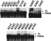
Cross-linking between P protein and PRNA. Cross-linking of B. subtilis RNase P holoenzyme reconstituted with equimolar-radiolabeled PRNA, pre-tRNA and AzP-labeled single-cysteine P protein variants (Niranjanakumari et al. 1998b). Lower mobility bands indicative of P protein–PRNA cross-linked species are indicated (XL).
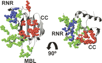
Summary of cross-linking and AOP cleavage experiments. Ribbon diagram of the B. subtilis P protein (Stams et al. 1998) showing regions where AzP- or AOP-modified single-cysteines cross-link to or cleave PRNA and pre-tRNA (Table 2). Highlighted amino acids indicate positions where modification results in cross-linking/cleavage of PRNA only (green), pre-tRNA only (red), or both pre-tRNA and PRNA (blue). Labels indicate the location of the RNR motif (RNR), metal binding loop (MBL) and central cleft (CC).
Cross-linking and cleavage of pre-tRNA and PRNA produced by incubation of the RNase P holoenzyme•pre-tRNA containing labeled P proteina
Probing PRNA–P protein interaction by hydroxyl-radical cleavage
While these cross-linking studies demonstrate that the RNR motif, N terminus, and metal binding loop of P protein are proximal to PRNA, identification of PRNA nucleotides involved in the cross-links is complicated by instability of PRNA during isolation of cross-linked species, leading to frequent termination bands in primer extension mapping experiments (data not shown). To overcome this limitation, we applied directed hydroxyl-radical cleavage to map the regions of PRNA near P protein residues (Sigman and Chen 1990; Huber 1993). For these experiments, we used single-cysteine P protein mutants derivatized with acetamido-1,10-o-phenanthroline (AOP), which upon addition of copper results in a covalently attached copper(II)-phenanthroline (Cu-OP) moiety. In the presence of mercaptopropionic acid, Cu-OP generates localized hydroxyl-radicals, cleaving proximal RNA regions. However, unlike Fe(II)–EDTA, which produces diffusible radicals, Cu-OP cleaves nucleic acid backbones through direct contact, preferably in the minor groove and in loops (Sigman and Chen 1990, and references therein). Thus, the effective radius between AOP-mediated cleavages and the point of attachment to the protein is primarily due to the length of the linker, and is relatively short (10–15 Å) compared to Fe(II)–EDTA (>24 Å) (Sigman and Chen 1990; Huq et al. 1999). AOP modification experiments therefore facilitate identification of specific PRNA nucleotides located near a predefined P protein amino acid in the RNase P•pre-tRNA complex, and provide important distance constraints complementary to photocrosslinking data.
As shown in Figure 4A, AOP modifications in the metal binding loop (Q38C, E40C, and D42C) lead to cleavage of PRNA at several positions. No cleavages at these nucleotide locations were observed using unmodified P protein or in the absence of copper and/or mercaptopropionic acid (data not shown); thus, the observed cleavage bands are specific to the covalently attached AOP moiety. Cleavage positions were mapped by comparison to RNase T1, RNase U2, and hydroxide cleavage ladders (Fig. 4A). AOP conjugation to each of the residues in the metal binding loop results in a prominent cleavage at nucleotide G34 and multiple nearby cleavages in the L3 loop (Fig. 4A; Table 2). Additionally, AOP incorporation at these positions produces cleavages near the 3′ end of PRNA that were mapped using 3′-end-labeled RNA. AOP modification of Q38C P protein leads to cleavage at nucleotides 320–321 in J18/2 and 359–360 near the internal bulge of P19. AOP-modified D42C P protein cleaves at nucleotides 355–356 of P19 (Fig. 4B; Table 2). These results indicate that the metal binding loop is <15 Å from L3, J18/2, and P19 in the RNase P•pre-tRNA complex.
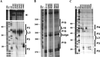
P protein/pre-tRNAAsp cross-linking and affinity cleavage. (A) Cleavage of PRNA by P protein modified with AOP in the metal binding loop or β-strand 3 in the RNaseP•pre-tRNA complex. Helix P2 and the L3 loop are marked. The arrow denotes cleavages near the 3′ end of PRNA. Control lanes show base hydrolysis, RNase T1, and RNase U2 ladders. Nucleotide positions are numbered. (B) AOP-mediated cleavages near the 3′ end of PRNA using 3′-end labeled PRNA. (C) PRNA cleavage by P protein with an AOP modification in the RNR motif. Helix P4, J3/4, and J2/3 are denoted. Lanes 10 (PRNA) and 11 (–) show cleavage of PRNA in the absence of P protein and in the presence or absence of mercaptopropionic acid, respectively.
Hydroxyl-radical cleavage assays were also performed to identify PRNA nucleotides proximal to the N terminus and β-strand 3 of P protein. As listed in Table 2, RNase P containing AOP linked to H3C cleaves PRNA prominently at nucleotides 26–28, corresponding to the 5′ side of helix P3 and the L3 loop. Modifications at residues K5C, R7C, K11C, or K12C also cleave PRNA in L3 (Table 2), indicating that the N terminus is near P3 in the RNase P•pre-tRNAAsp complex. Modification of amino acids in β-strand 3 promotes cleavage in several regions of PRNA. AOP modification at R45C causes cleavages mainly in L3, similar to modifications in the metal binding loop (Fig. 4A; Table 2), while modification at residue 46 cleaves predominately at nucleotide 21 in helix P2, positioning β-strand 3 near the J2/3 region of PRNA.
AOP cleavage analysis also indicates that the conserved RNR motif is located close to helix P4, a highly conserved RNA structure thought to be part of the active site in the RNase P•pre-tRNA complex (Fig. 4C; Table 2; Brown et al. 1991; Hardt et al. 1995b, 1996; Harris and Pace 1995; Christian et al. 2002a; Crary et al. 2002; Xiao et al. 2002). Modification of R60C results in a prominent cleavage at nucleotide A49. Modification of K64C leads to PRNA cleavages at A48, A49, and G50 in helix P4. In addition, modifications in the RNR motif result in cleavages in structural features near P4. Modification at N61C cleaves PRNA at nucleotides 43–49 in J3/4 and P4, and 21–22 in P3. In addition, AOP-modified R62C P protein cleaves mainly at G43 in J3/4 while AOP-modified R65C and R68C P protein cleave at nucleotides 21–23 in P2 and J2/3. Finally, AOP modifications at R62C and K64C cause cleavages near the 3′ end of PRNA at nucleotides 366–367 of J19/4 (AOP-R62C P protein) and 370–371 of P19 stem (AOP-R64C P protein). Together, these data indicate that the RNR motif is located close to the catalytic core of RNase P RNA. This observation, together with the strong conservation of the RNR motif, suggests that this region plays a fundamental role in RNase P function.
Previous biochemical studies have shown that the B. subtilis P protein can functionally substitute for the protein component of Escherichia coli RNase P both in vitro and in vivo (Guerrier-Takada et al. 1983; Wegscheid et al. 2006). Moreover, recent biochemical studies of the RNase P holoenzyme in the absence of substrate have suggested that the positioning of P protein is likely to be similar in these heterologous holoenzymes (Buck et al. 2005a,b). To further test this hypothesis, and examine the consistency of P protein positioning in the presence of the substrate, we examined the AOP footprinting of B. subtilis single-cysteine P proteins in combination with E. coli RNase P RNA (M1 RNA). In general, we observed similar AOP-mediated cleavages of B. subtilis and E. coli RNase P RNAs when complexed with pre-tRNAAsp (cf. Fig. 1 and Fig. 5). For M1 RNA, metal binding loop variants Q38C, E40C, and D42C produce strong cleavages around G51 and G52 on the 3′ side of P3, and minor cleavages at positions 29–33 on the 5′ side of the P3 (Fig. 5). Thus, the metal binding loop is proximal to P3 in both enzyme–substrate complexes. In β-strand 3, AOP-labeled V46C cleaves M1 RNA at nucleotides 18–19 in the helix P2, indicating a location near J2/3 as seen in the B. subtilis holoenzyme complex. Similarly, the AOP-derivatized single-cysteines in the RNR motif cleave M1 RNA in helix P4. Modification at positions R60C and K64C produce cleavages at nucleotides 67–69 on the 5′ side of P4 while modification of N61C protein causes cleavages at positions 63–68 in J3/4 and P4. Once again, these results are comparable to the observed cleavages in B. subtilis holoenzyme, indicating that the position and orientation of P protein is similar in both enzyme–substrate complexes.
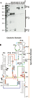
Affinity cleavage of E. coli PRNA by AOP-modified B. subtilis P protein. Cleavage of M1 RNA by AOP-modified P protein in the presence of B. subtilis pre-tRNAAsp. (A) Cleavages in the region from P1 through P4; J2/3, P3, and P4 are marked. Control lanes show base hydrolysis (OH), RNase T1, and RNase U2 ladders, and a control reaction in the presence of wild-type P protein. Nucleotide positions are numbered. (B) Secondary structure of M1 RNA catalytic domain drawn according to the topology of the T. maritima crystal structure (Frank and Pace 1998; Torres-Larios et al. 2005), indicating cleavage sites for AOP-labeled single-cysteine P protein variants: (red arrow) RNR motif; (black arrow) metal binding loop; and (black diamond) β-strand 3. Shaded arrows denote strong cleavage bands; open arrows denote weaker cleavage bands. Highlighted nucleotides indicate the effect of the protein component on Fe–EDTA cleavage of PRNA, including site protection from cleavage (red), enhancement of cleavage (blue), and variable effect (green) (Buck et al. 2005b).
Regions of P protein that are near pre-tRNA
Cross-linking of P protein variants to pre-tRNA
Previously, we demonstrated that azidophenacyl moieties attached to cysteine residues in the central cleft (F16, F20, V32, Y34, S49, and I86) of P protein cross-link to the pre-tRNA leader in the RNase P•pre-tRNA complex (Niranjanakumari et al. 1998b). To further determine the location and orientation of P protein in the enzyme–substrate complex, we identified additional P protein amino acids that cross-link to pre-tRNA substrates. Cross-linking experiments were carried out using 5′-radiolabeled pre-tRNAAsp and 10 mM Ca2+, as described above for PRNA cross-linking.
Cross-linked pre-tRNA•P protein species were observed for modified cysteines located in β-strand 1 (S25C); the RNR motif (S51C, K52C, and K53C); and β-strand 4 (R88C and R89C; data not shown; Fig. 3). No cross-links were observed for AzP- modification at the N terminus (H3C, K5C, R7C, R9C, K11C, K12C, and D15C), metal binding loop (Q38C, E40C, and D42C), β-strand 3 (R45C and V46C), and the RNR motif (A57C, N61C, R62C, and R65C). Together, these results indicate that both the RNR motif and central cleft of P protein are proximal to substrates and further constrain the location and orientation in the enzyme–substrate complex.
Affinity cleavage of pre-tRNA by P protein variants
We also probed the local structural environment of residues in the central cleft and the RNR motif using AOP modification. In the central cleft, we examined cleavages due to modification of residues located on helix A and β-strand 4, which comprise the central cleft. As predicted from previous cross-linking experiments (Niranjanakumari et al. 1998b), AOP modifications at these positions cleave the pre-tRNA leader at nucleotides –3 to –7 (Fig. 6A; Table 2). These cleavage data confirm that the central cleft of P protein is located near the leader sequence of pre-tRNA in the holoenzyme•substrate complex, and demonstrate that AOP experiments provide similar data to the sites of photocrosslinks mapped using reverse transcription (Niranjanakumari et al. 1998b).
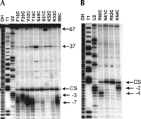
Affinity cleavage of pre-tRNAAsp by AOP-modified P protein. (A) Cleavage of radiolabeled pre-tRNA by RNase P formed with P protein modified with AOP in the central cleft. Control lanes show base hydrolysis (OH), RNase T1, and RNase U2 ladders. The RNase P cleavage site (CS) in pre-tRNA, 5′ leader nucleotides –3 and –7, and nucleotides 37 and 67 are labeled. (B) Cleavage of pre-tRNA by RNase P formed with P protein modified with AOP in the RNR motif. Control lanes are as in A. The RNase P cleavage site (CS) and 5′ leader positions–2 and –4 are shown.
In the RNR motif, AOP modifications at residues R60C and K64C produce major cleavage bands at the –4 and –2 positions, respectively (Fig. 6B). These assays were performed in the presence of CaCl2 to minimize enzymatic cleavage of pre-tRNA; however, a small fraction of substrate was converted to mature tRNA in all of experiments (Fig. 6). The intensity of the band at the pre-tRNA cleavage site is increased in experiments examining cleavage of PRNA complexed with AOP-labeled N61C or R62C P proteins, when compared to that of R60C and K64C (Fig. 6B). Since the cleavage rate constants for holoenzymes containing these AOP modifications are comparable in the absence of Cu (Table 1), these data suggest that AOP modifications at N61C and R62C may be located near the pre-tRNA cleavage site. AOP modifications in the RNR motif also cleave pre-tRNA near the 3′ end (Fig. 6A) at nucleotides G65, U66, and C67. These nucleotides are located at the base of the acceptor stem, 8 nt on the 5′ side of the 3′-terminal RCCA motif (Table 2).
In summary, these data indicate that the RNR motif is located proximal to and on the 5′ side of the pre-tRNA cleavage site. Strikingly, AOP modifications in the RNR motif cleave both PRNA helix P4 and near the cleavage site of bound pre-tRNA (Figs. 4C, 6B; Table 2). These results confirm that the RNR motif of the protein, helix P4 of PRNA, and the cleavage site of pre-tRNA are all located in the catalytic core of the RNase P holoenzyme, and that the active site of RNase P holoenzyme is near the RNA–protein interface.
Holoenzyme structure modeling
The experiments described above not only reveal regions of PRNA and pre-tRNA that are proximal to P protein in the holoenzyme–substrate complex, but also provide detailed distance constraints useful in modeling the holoenzyme–substrate complex. Previously, low-resolution tertiary structure models of bacterial PRNA–substrate and holoenzyme–substrate complexes have been derived from phylogenetic and biochemical data (Westhof and Altman 1994; Chen et al. 1998; Massire et al. 1998; Tsai et al. 2003). Given that the experiments described here probe the solution structure of the RNase P complex, they are more directly comparable with these low-resolution biochemical models and the data upon which they are based than to recent X-ray crystal structures of PRNA. We therefore undertook modeling experiments beginning from a published model of B. subtilis PRNA based on biochemical data (Massire et al. 1998), rather than on a crystal structure.
The position of P protein with respect to PRNA and pre-tRNA (containing a 9-nt 5′ leader) was modeled using the program CNS (Brunger et al. 1998). Structure calculations were performed starting with the X-ray crystal structure of the P protein (Stams et al. 1998) and a model of the PRNA•pre-tRNA complex (Massire et al. 1998) containing nucleotides 4–395 of the PRNA and a pre-tRNA molecule that includes nucleotides −9 to 72. To minimize changes in the structure of each individual component of the complex during the modeling procedure, an extensive set of intramolecular restraints was generated based on the coordinates of the starting structures. Additionally, the hydroxyl-radical cleavage data (as annotated in Table 2) were translated into restraints for CNS by defining each cleavage as a distance linking the C1′ atom of an RNA nucleotide with the Cα atom of the corresponding protein residue. This assignment was based on the ability of AOP to cleave DNA by removing hydrogen from either the C1′ or C4′ positions with a preference for attack at the C1′ in double-stranded DNA (Sigman and Chen 1990).
Using rigid-body minimization, an initial family of structures was generated that satisfied all distance constraints with the exception of those derived from AOP cleavages in P19. P19 is not well constrained in initial structural models, as these structures were generated using comparative sequence analysis (Massire et al. 1998) or cross-linking and footprinting experiments using only E. coli M1 RNA, which lacks helix P19 (Harris et al. 1997; Chen et al. 1998; Tsai et al. 2003). In addition, the region of P19 cleaved by AOP-modified P proteins is not resolved in the crystal structure of Bacillus stearothermophilus PRNA (Kazantsev et al. 2005). Structures were therefore regenerated without constraints involving P19. The structure shown in Figure 7 depicts the representative from this family of structures with the lowest residual energy and no distance or dihedral angle restraint violations. When the protein structure was extracted from the complex and compared against the starting crystal structure, a backbone RMSD of 0.21 Å was obtained. Similarly, the PRNA and pre-tRNA structures extracted from the model shown in Figure 7 agree well with their starting structures, giving backbone RMSDs of 1.01 Å and 0.81 Å, respectively. This model places key regions of P protein in close proximity to functionally important regions of PRNA and pre-tRNA. Specifically, the RNR motif is proximal to PRNA helix P4 as well as the acceptor stem of pre-tRNA, the central cleft of the P protein interacts with the leader sequence of pre-tRNA, and the metal binding loop and N terminus of P protein are proximal to the P3 stem–loop. In addition, the pre-tRNA cleavage site is located proximally to both P4 and the RNR motif of the P protein.
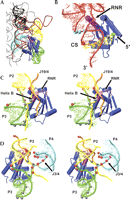
Model of B. subtilis PRNA•P protein•pre-tRNA ternary complex. (A) Holoenzyme substrate model based on affinity cleavage results. tRNA is shown in red, and relevant PRNA structures are highlighted: P1 (black), P2 (yellow), J2/3 and P3 (green), P4 (cyan). P protein is shown in blue with the RNR motif highlighted in magenta. (B) Detail of the predicted P protein•pre-tRNA interface. RNA and protein colored as in A, with the pre-tRNA 5′ leader shown in yellow. The RNase P cleavage site is marked with a red sphere. AOP cleavage sites (Table 2) are marked with spheres: RNR motif (cyan), RNR motif and central cleft (gray), central cleft (green). (C) Stereo image of the predicted P protein•PRNA interface. RNA and protein are colored as in A, with J19/4 shown in orange. AOP cleavage sites are marked with spheres: RNR motif (red), metal binding loop (gray). (D) Alternate perspective of C showing the protein location relative to P4 and J3/4 (cyan).
DISCUSSION
Despite its relatively simple composition of a single protein and single RNA, in bacterial RNase P interactions between protein and RNA subunits synergistically give rise to function. In this complex, P protein modulates many aspects of RNase P function, including PRNA structure (Buck et al. 2005a, 2005b), metal affinity (Kurz and Fierke 2002), cleavage site selection (Guerrier-Takada et al. 1989; Zahler et al. 2005), catalysis (Kurz et al. 1998; Sun et al. 2006), and substrate affinity through contacts to both PRNA and to the 5′-leader sequence of substrates (Crary et al. 1998; Niranjanakumari et al. 1998b; Day-Storms et al. 2004; Rueda et al. 2005). Given the complex function of the protein subunit and its apparent proximity to the RNase P active site, it is of great interest to understand its positioning and orientation within the enzyme–substrate complex and to identify protein side chains that are involved in specific intermolecular interactions with PRNA and pre-tRNA.
While structural models of the RNase P holoenzyme–substrate complex based on biochemical and X-ray crystallographic data have been previously proposed, the positioning and orientation of P protein within these models is poorly constrained (Christian et al. 2002b; Harris and Christian 2003). Based upon cross-linking, Fe(II)–EDTA cleavage, and the leader length dependence of substrate binding, most previous models have positioned P protein such that the face proximal to PRNA is located near N(−5) of pre-tRNA, ∼20 Å from the cleavage site (Niranjanakumari et al. 1998b; Tsai et al. 2003; Buck et al. 2005b). In contrast, recent data from time-resolved FRET distance measurements indicate that positions −4 through −7 of the pre-tRNA leader interact with P protein (Rueda et al. 2005). These data position P protein closer to the pre-tRNA cleavage site and the catalytic core of PRNA than previously proposed, suggesting that P protein may form an integral part of the active site.
In the present investigation, we used a combination of site-specific single-cysteine mutations, photocrosslinking, and covalently attached copper phenanthroline (AOP)-mediated affinity cleavage assays to probe the protein–RNA interfaces in the RNase P holoenzyme. Unlike Fe(II)–EDTA, AOP cleavage does not proceed through a diffusible hydroxyl-radical (Sigman and Chen 1990; Pan et al. 1994; Papavassiliou 1995); however, several caveats apply to the interpretation of AOP cleavage experiments. The reactive conformation of the nucleic acid may be infrequently observed and not represent the average conformation. In addition, while not all reactive sites must be single stranded, AOP cleavage is influenced by secondary structure, with a preference for single-stranded nucleic acids (Huber 1993). Nonetheless, AOP cleavage offers the important advantage of providing firm distance constraints between specific protein amino acids and RNA nucleotides that are usable in the development of structural models.
Based on our cross-linking and AOP cleavage experiments, we developed a structural model of the RNase P enzyme–substrate complex with improved resolution for the positioning of P protein. For this analysis we began with a previously published model of the PRNA–pre-tRNA complex based on biochemical and phylogenetic data (Massire et al. 1998). Use of this biochemical model allows for the comparison and incorporation of our distance constraint data with the large body of similar data from previous biochemical investigations of RNase P structure in solution. In addition, use of the model avoids complications introduced by unresolved regions and the effects of crystal packing contacts evident in the catalytic domains of both PRNA crystal structures (Kazantsev et al. 2005; Torres-Larios et al. 2005). While the biochemical model differs in detail to recently published crystal structures of PRNA, the topology and relative orientation of RNA helices of interest for this study (P1–P4) are similar (Fig. 8). We have mapped our affinity cleavage data from B. subtilis RNase P onto the crystal structure of PRNA from the low G/C Gram-positive bacterium B. stearothermophilus (Kazantsev et al. 2005). Comparison of the relevant portions of the two RNA structures (P1–P4, J18/2 and J19/4) suggests that, within the resolution of our current model (∼10 Å), the structural model and crystal structure would yield comparable results (Fig. 8). Importantly, the final model shown in Figure 7 is consistent with all our cleavage results, except those observed in P19. These cleavages could be explained by either transient dimer formation, a significant change in the conformation of P19 in the presence of P protein, or conformational flexibility in the holoenzyme–substrate complex. In addition, several AOP cleavages appear in regions of PRNA that, while proximal to the protein, do not appear to be accessible in the model due to intervening RNA structures. This is especially notable for J19/4, which appears to intercede between the RNR motif and several associated AOP cleavage sites in J3/4 and P4. The presence of such cleavages could also be explained by changes in the positioning of these regions upon pre-tRNA binding or conformational dynamics.
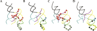
Locations of AOP cleavage sites on PRNA sites of AOP cleavage are shown mapped onto a biochemical model of B. subtilis PRNA (Massire et al. 1998; (A,B) and X-ray crystal structure of B. sterothermophilus PRNA (Kazantsev et al. 2005; (C,D). Regions of secondary structure containing the majority of cleavage sites are shown: P1 (black), P2 and J2/3 and J18/2 (yellow), P3 (green), P4 and J3/4 (cyan), and J19/4 (violet). Cleavage sites from regions of P protein (Table 2) are marked with spheres: (A,C) RNR motif (red), metal binding loop (gray), (B,D) N terminus (yellow), β-strand 3 (blue), and both the N terminus and β-strand 3 (black).
Perhaps the most striking result of the modeling experiments shown in Figure 7 is the proximity of the RNR motif to PRNA helix P4 and the RNase P active site (Fig. 7). Helix P4 contains 11 out of the 21 invariant nucleotides in RNase PRNA (Frank and Pace 1998), along with base and backbone functional groups important for binding and catalysis (Hardt et al. 1995a, 1996; Harris and Pace 1995; Kazantsev and Pace 1998; Heide et al. 1999; Siew et al. 1999). Moreover, phosphorothioate substitution experiments have suggested that P4 is important for positioning divalent metal ions required for catalysis (Christian et al. 2000, 2002b; Crary et al. 2002). AOP-mediated cleavages indicate that the RNR motif of P protein is proximal to P4, positions –2 and –4 of pre-tRNA, and potentially the pre-tRNA cleavage site (Table 2). In addition, the AOP modifications at position K52 indicate that the RNR motif is proximal to the base of the pre-tRNA acceptor stem. This observation is consistent with recent short-range cross-linking results placing the universally conserved bulged uridine in P4 in close proximity to the pre-tRNA acceptor stem (Christian et al. 2006).
Given the highly basic nature of the RNR motif and its proximity to helix P4 and the cleavage site, we propose that this conserved motif may be positioned to stabilize binding interactions between pre-tRNA and/or catalytically important metal ions and P4. Based on the results from the present investigation, it is tempting to speculate that helix B of P protein may interact with the major groove of P4. The major groove of this helix may be widened sufficiently by the two bulged nucleotides suggested by the crystal structure of B. stearothermophilus PRNA to accommodate such an interaction (Kazantsev et al. 2005). A similar mode of interaction has been observed between the arginine-rich peptide of HIV-1 Rev protein and double-stranded RNA (Battiste et al. 1996). It is possible that the conserved sequence and structures of P4 and the RNR motif function cooperatively to stabilize the active site structure of the RNase P holoenzyme and/or the holoenzyme–substrate complex.
Modeling based on AOP cleavage experiments also suggests the possibility of multiple regions of interaction between P protein and pre-tRNA substrates. In agreement with previous results (Crary et al. 1998; Niranjanakumari et al. 1998b; Day-Storms et al. 2004; Rueda et al. 2005), the data presented here indicate that the central cleft of P protein is proximal to the pre-tRNA leader and not PRNA. Pre-tRNA cleavages are also observed for AOP modifications in the RNR motif and β-strand 4. Consistent with these observations, the model shown in Figure 7 positions conserved residues in the RNR motif near the base of the pre-tRNA acceptor stem (Fig. 7B). This proximity, together with the recently observed ability of P protein to alter mature tRNA binding to the RNase P holoenzyme (Sun et al. 2006), suggests the possibility of additional P protein/pre-tRNA contacts in this region.
The data presented here also emphasize the importance of helix P3 for P protein binding. In our model, P3 is in close proximity to both the N terminus and metal binding loop of P protein, two P protein features that have been suggested as possible RNA binding motifs (Stams et al. 1998). In addition, β-strand 3, which includes part of the RNR motif, is located near L3, P3, and J3/4 (Fig. 7C). The minimal consensus structure of the bacterial PRNA retains P3, although no highly conserved nucleotides are present in this helix (Frank and Pace 1998). A homolog of the P3 stem–loop is also identifiable in archaeal and eukaryotic RNase PRNA, although these structures can differ significantly in primary sequence. In Eukaryotes, the structure of helix P3 is elaborated considerably relative to the Archaeal and Bacterial structures, suggesting that a more complex functional role for this helix, perhaps in binding of one or more eukaryotic protein components (Ziehler et al. 2001). In the absence of P protein, 4-thiouridine incorporated at the N(–10) position of the pre-tRNA leader cross-links to P3 of E. coli M1 RNA (Christian and Harris 1999). In our model of the RNase P holoenzyme, P protein is positioned between the pre-tRNA leader and helix P3. Thus, the 5′ leader of pre-tRNA may adopt a similar conformation in both the holoenzyme and PRNA alone, with P3 playing a role in protein, rather than substrate, binding.
The model of P protein bound to PRNA shown in Figure 7 is also consistent with data from numerous previous studies. Figure 1 shows that the regions suggested to be involved in protein binding by our model are coincident with those consistently protected from cleavage by the presence of P protein (Buck et al. 2005b). Also, much of P3 and L3, while implicated in protein binding, remain solvent exposed in this model, in keeping with the absence of protections in these regions. However, the regions where P protein binding leads to enhancement of Fe(II)–EDTA, cleavages in PRNA do not overlap with AOP-mediated cleavages (e.g., J18/2, L15; Fig. 1B), suggesting that these changes reflect alterations in the PRNA conformation. Similarly, this holoenzyme model is consistent with the observed cleavages in J18/2 of E. coli PRNA caused by Fe(II)–EDTA covalently attached to E. coli C5 protein (Biswas et al. 2000; Tsai et al. 2003). Finally, like previous reports, our results indicate that P protein binds in a similar position in a heterologous holoenzyme formed with E. coli M1 RNA, and suggest that this model is applicable to bacterial RNase P in general.
MATERIALS AND METHODS
Chemicals and buffers
Chemicals of the highest purity were purchased. Nucleotide triphosphates and cytosine monophosphate were purchased from Amersham BioScience. Unless specified otherwise, calf intestinal alkaline phosphatase, T4 polynucleotide kinase, and restriction enzymes were purchased from New England Biolab. Azidophenacyl (AzP) bromide was purchased from Sigma, and 5-iodoacetamido-1,10-o-phenanthroline was purchased from Molecular Probes. All buffers were made using water deionized by a Milli-Q system (Milliport Corp.). Single turnover experiments were performed in Buffer A (10 mM MgCl2, 400 mM NH4Cl, and 50 mM MES-Tris buffer, pH 6.0) at 37°C. Affinity cleavage experiments were performed in the same buffer conditions, but MgCl2 was replaced with CaCl2 (Buffer B). Cross-linking experiments were performed in Buffer C (10 mM CaCl2, 100 mM NH4Cl, and 50 mM MES/50 mM Tris, pH 6.0, at 37°C).
RNA and protein preparation
B. subtilis PRNA, E. coli M1 RNA, and B. subtilis pre-tRNAAsp containing a 14 nt leader sequence were prepared by in vitro transcription from linearized plasmids using T7 RNA polymerase (Milligan and Uhlenbeck 1989). PRNA and pre-tRNA were 5′-end-labeled with γ-32P-ATP and T4 polynucleotide kinase, and purified by denaturing polyacrylamide gel electrophoresis (7 M urea and 8% polyacrylamide) as described (Kurz et al. 1998). For 3′-end labeling, RNA was radioactively labeled using standard protocols (Perrotta and Been 1990). [5′-32P]pCp was prepared by incubating CMP (40 μM) with T4 polynucleotide kinase in 4 μM γ-32P-ATP, 50 mM Tris-HCl, pH 8.0, 10 mM MgCl2, and 15 mM dithiothreitol (DTT) at 37°C for 30 min. The reaction was terminated by heating to 95°C for 5 min. Subsequently, PRNA (3 μM) was labeled by incubation with 5 μM [5′-32P]pCp, 0.1 mM ATP, 50 mM HEPES, pH 7.5, 3.3 mM DTT, 15 mM MgCl2, and 10% DMSO in the presence of T4 RNA ligase (50 units) at 37°C for 2 h. The labeled RNA was gel purified, eluted, and further purified by ethanol precipitation followed by gel filtration over a G-25 Sephadex centrifuge column.
Variants of P protein with site-specific cysteine mutations were prepared as described previously (Niranjanakumari et al. 1998a) and subcloned into pET28b (Novagen). The recombinant single-cysteine P proteins were expressed in BL21(DE3)pLysS E. coli by growth at 37°C to an OD600 of 0.6–0.8, induction with isopropylthio-β-D-galactopyranoside (0.4 mM), and incubation at 37°C for 4 h (Niranjanakumari et al. 1998a). Proteins were purified from the soluble fraction of the cell lysate by CM-Sepharose ion-exchange chromatography in the presence of urea, and the concentration was determined by measuring the absorbance at 280 nm (ε280 = 5120 M−1cm−1) (Niranjanakumari et al. 1998a). Recombinant E. coli C5 protein with a hexahistadine tag was prepared as previously described (Riveraleon et al. 1995).
RNAs were refolded immediately prior to use by incubation at 95°C for 3 min, followed by a twofold dilution into reaction buffer (2× Buffer A, B, or C) and incubation at 37°C for 15 min. Holoenzyme was reconstituted immediately prior to use by incubation of refolded PRNA with an equimolar concentration of P protein at 37°C for 10 min in the appropriate reaction buffer.
Derivatization of P protein cysteine variants
Single-cysteine P protein variants (100 μM) were derivatized by incubation with either AzP (700 μM) or 5-iodoacetamide-1,10-o-phenanthroline (1 mM) at room temperature for 3 h in the dark in 50 mM Tris-HCl, pH 7.5, 100 mM NaCl, 5% glycerol, 1 mM tris(2-carboxyethyl)phosphine. Derivatized protein was separated from excess reagent using a PD-10 gel filtration column (Pharmacia). The extent of modification was >95%, as determined by reaction of the residual free thiol group with 5,5′-dithio-bis(2-nitrobenzoic acid) (Sigma) according to standard protocols (Creighton 1990).
Single-turnover experiments
Single-turnover kinetic assays were performed using excess RNase P holoenzyme (E, 400 nM) and limiting 32P-labeled pre-tRNAAsp (<0.1 nM) containing a 14-nucleotide leader sequence in Buffer A (10 mM MgCl2, 400 mM NH4Cl, and 50 mM MES-Tris buffer, pH 6.0) at 37°C as previously described (Beebe and Fierke 1994). Under these conditions the observed single turnover rate constant is not dependent on [E] (data not shown). Reactions were quenched by dilution into an equal volume of 100 mM EDTA (pH 8.0), 0.05% (w/v) bromophenol blue, 0.05% (w/v) xylene cyanol, and 8 M urea; 5′ -end-labeled precursor and product were separated by 8% PAGE (Beebe and Fierke 1994), and the fraction cleaved was determined using a PhosphorImager (Amersham Bioscience Corp.). The observed rate constant was calculated from fitting a single-exponential decay to these data using Kaleidagraph software (Synergy Software).
Cross-linking assays
RNase P holoenzyme cross-linking was performed by reconstituting the holoenzyme with equimolar concentrations of PRNA and an AzP-derivatized P protein variant (20 nM each) in the presence of excess pre-tRNAAsp substrate (100 nM) in Buffer C; 5′-end-labeled PRNA was used to probe PRNA-P protein interactions, while 5′-end-labeled B. subtilis precursor tRNAAsp with a 14 nt leader (Crary et al. 1998) was used to probe pre-tRNA–P protein interactions. After incubating at 37°C for 20 min, samples were irradiated for 1 min on ice using a 312-nm hand-held UV lamp at ∼5 cm distance with a polystyrene filter. Samples were denatured in SDS-sample buffer (50 mM Tris-Cl, pH 6.8, 0.25% SDS, 0.1 M DTT, 10% glycerol) by heating at 85°C for 5 min and analyzed by 7.5% or 15% SDS-PAGE.
Affinity cleavage assays
Affinity cleavage experiments were performed with either radiolabeled B. subtilis P RNA, E. coli M1 RNA, or pre-tRNA to probe RNase P RNA or pre-tRNA contacts. RNase P holoenzyme was reconstituted by incubating equimolar concentrations of PRNA and AOP-modified P protein (0.4 μM each) in Buffer B at 37°C for 10 min. Pre-tRNA substrate was then added and incubated for 20 min before the addition of CuSO4 (final concentration, 50 μM). After incubating for 10 min, the reaction was initiated by adding mercaptopropionic acid (3 mM), incubated for 30 min at 37°C, and stopped by addition of neocuproine (1 mM). Nucleic acids were precipitated with 2.5 volumes of ice-cold ethanol, and washed twice with 70% ethanol. The dried samples were resuspended in urea-dye solution (4 M urea, 50 mM EDTA, traces of bromophenol blue, and xylene cyanol) and analyzed on 8%–15% polyacrylamide-urea gels. The gels were dried and scanned using a PhosphorImager (Amersham Bioscience Corp.), and cleavage sites were identified by comparison to an alkaline hydrolysis ladder, RNase T1, and RNase U2 digests.
Structure modeling
Holoenzyme–substrate models were generated using version 1.0 of the program CNS (Brunger et al. 1998). Structure calculations were performed starting with the X-ray crystal structure of the P protein (Stams et al. 1998) to which protons were added using standard protocols. For RNA components, a model of the PRNA–pre-tRNAPhe complex, described by Westhof and coworkers (Massire et al. 1998), was used after conversion to a CNS compatible format followed by the addition of protons. This PRNA–pre-tRNA model contains nucleotides 4–395 of the RNA and a pre-tRNA molecule that includes nucleotides −9 to 72. To minimize changes to the structure of each individual component of the complex during the docking procedure, an extensive set of intramolecular restraints was generated based on the coordinates of the starting structures. From the crystal structure of the RNase P protein, restraint lists were generated involving all NH–NH, C–Cα, and NH–Cα distances <12 Å. In a similar way, backbone dihedral angles were extracted and used as input to the CNS refinement protocol. For the RNA molecules, restraint lists were generated involving all N3–C4′, N3–N3, and C4′–C4′ distances <12 Å. Additionally, the hydroxyl-radical cleavage data (Table 2) were translated into restraints for CNS by defining each cleavage as a distance linking the C1′ atom of a PRNA or pre-tRNA nucleotide with the Cα atom of the corresponding protein residue. A lower bound of 1.8 Å and an upper bound of 20 Å were used for each of these restraints.
Structures were generated using rigid-body minimization. This minimization procedure involves a low-temperature torsion angle dynamics simulated annealing stage followed by Powell minimization (Chou et al. 2000; Goto et al. 2001). While performing molecular dynamics in torsion angle space, the RNA and pre-tRNA structures were held fixed while the protein was allowed to translate and rotate. All of the molecules were free to move during the Powell minimization stage. The torsion angle dynamics stage included cooling from 200 to 0 K in 5-K steps. At each temperature, 15 psec of molecular dynamics were performed using a 15-psec time step. Backbone dihedral angles were restrained with a force constant of 200 kcal/mol with an acceptance angle of ±0.1° with respect to the input target value. Distances were restrained with a 150 kcal/mol force constant with a ±0.2 Å acceptance region. During the cooling process, the van der Waals force constant was increased from 0.1 to 1 kcal/mol Å2. Following the torsion angle dynamics stage, the structures were furthered refined with 10 cycles of Powell minimization. Each cycle contained 2000 minimization steps. During this stage, the dihedral angle force constant was 300 kcal/mol and the distance force constant was 150 kcal/mol.
ACKNOWLEDGMENTS
We thank Manoj Cheriyan for his help in creating the figures, Lance Rider for his assistance, and Dr. Marcy Hernick and Dr. James Hougland for their comments. This research was supported by National Institutes of Health (NIH) Grant GM55387. Partial support was provided by NIH Training Grants GM08487 and GM008270 (J.J.D.). N.H.Z. was partially funded by NIH postdoctoral fellowship F32ES013881.
Footnotes
-
↵3 Present address: Department of Natural Sciences and Mathematics, Southeastern University, 1000 Longfellow Boulevard, Lakeland, FL 33801, USA.
-
↵4 Department of Biochemistry and Cell Biology, Center for Structural Biology, State University of New York at Stony Brook, Stony Brook, NY 11794, USA.
-
Reprint requests to: Carol A. Fierke, Chemistry Department, University of Michigan, 930 N. University Avenue, Ann Arbor, MI 48109, USA; e-mail: fierke{at}umich.edu; fax: (734) 647-4865.
-
Abbreviations: RNP, ribonucleoprotein; pre-tRNA, precursor tRNA; PRNA, the RNA component of the B. subtilis RNase P holoenzyme; P protein, the protein component of the B. subtilis RNase P holoenzyme; RNase P, ribonuclease P; Cu-OP, copper 1,10-o-phenanthroline; AOP, acetamido-1,10-o-phenanthroline; M1 RNA, the RNA component of the E. coli RNase P holoenzyme; C5 protein, the protein component of the E. coli RNase P holoenzyme; AzP, azidophenacyl moiety; C-domain, the PRNA catalytic domain; S-domain, the PRNA specificity domain; DTT, dithiothreitol; RNR, highly conserved region of the P protein consisting of arg/asn/arg; MBL, metal binding loop in the P protein; CC, central cleft of the P protein.
-
Article published online ahead of print. Article and publication date are at http://www.rnajournal.org/cgi/doi/10.1261/rna.308707.
-
- Received September 21, 2006.
- Accepted December 21, 2006.
- Copyright © 2007 RNA Society




