


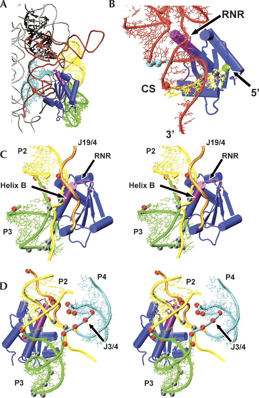
Model of B. subtilis PRNA•P protein•pre-tRNA ternary complex. (A) Holoenzyme substrate model based on affinity cleavage results. tRNA is shown in red, and relevant PRNA structures are highlighted: P1 (black), P2 (yellow), J2/3 and P3 (green), P4 (cyan). P protein is shown in blue with the RNR motif highlighted in magenta. (B) Detail of the predicted P protein•pre-tRNA interface. RNA and protein colored as in A, with the pre-tRNA 5′ leader shown in yellow. The RNase P cleavage site is marked with a red sphere. AOP cleavage sites (Table 2) are marked with spheres: RNR motif (cyan), RNR motif and central cleft (gray), central cleft (green). (C) Stereo image of the predicted P protein•PRNA interface. RNA and protein are colored as in A, with J19/4 shown in orange. AOP cleavage sites are marked with spheres: RNR motif (red), metal binding loop (gray). (D) Alternate perspective of C showing the protein location relative to P4 and J3/4 (cyan).




