


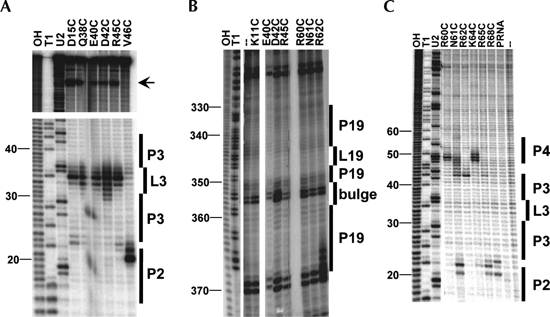
P protein/pre-tRNAAsp cross-linking and affinity cleavage. (A) Cleavage of PRNA by P protein modified with AOP in the metal binding loop or β-strand 3 in the RNaseP•pre-tRNA complex. Helix P2 and the L3 loop are marked. The arrow denotes cleavages near the 3′ end of PRNA. Control lanes show base hydrolysis, RNase T1, and RNase U2 ladders. Nucleotide positions are numbered. (B) AOP-mediated cleavages near the 3′ end of PRNA using 3′-end labeled PRNA. (C) PRNA cleavage by P protein with an AOP modification in the RNR motif. Helix P4, J3/4, and J2/3 are denoted. Lanes 10 (PRNA) and 11 (–) show cleavage of PRNA in the absence of P protein and in the presence or absence of mercaptopropionic acid, respectively.




