


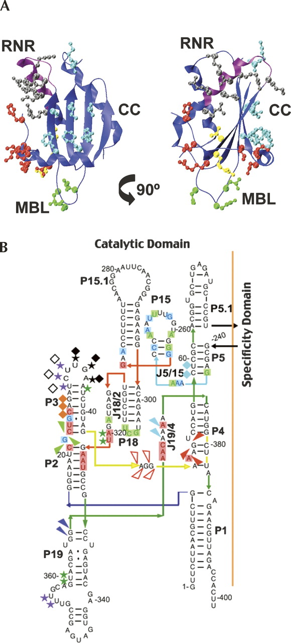
Structures of B. subtilis PRNA and B. subtilis P protein. (A) Structure of B. subtilis P protein indicating sites of single-cysteine mutations. The protein backbone is shown in blue with the RNR motif highlighted in magenta. Labels indicate the location of the RNR motif (RNR), metal binding loop (MBL), and central cleft (CC). Amino acids replaced with single-cysteine mutations near the N terminus (red), metal binding loop (green), RNR motif (black), β-strand 3 (yellow), and central cleft (cyan) are shown. (B) Secondary structure of the catalytic domain of B. subtilis PRNA, based on topology from the X-ray structure of the B. stearothermophilus PRNA (Kazantsev et al. 2005), indicating cleavage sites for AOP-labeled single-cysteine P protein variants: (arrow) RNR motif and β-strand 3 (red=R60, N61, K64; white=N61, N62; green=V46, N61, R65, R68; dark blue=R62; light blue=K64); (star) metal binding loop and β-strand 3 (black=Q38, E40, D42, R45; pink=Q38; purple=D42); and (diamond) N terminus (black=H3, K5, K7, K11, K12; white=H3, R7; orange=H3; aqua=K12) (summarized in Table 2). Shaded markers denote strong cleavage bands; open markers denote weaker cleavage bands. Highlighted nucleotides indicate sites where P protein reduced (red), enhances (blue), or has a variable effect (green) on cleavage during in-gel Fe(II)–EDTA cleavage assays (Buck et al. 2005b).




