


KRGG1 function in RNA editing in Trypanosoma brucei
- 1Seattle Children's Research Institute, Seattle, Washington 98109, USA
- 2Department of Pediatrics and Global Health, University of Washington, Seattle, Washington 98105, USA
- Corresponding author: ken.stuart{at}seattlechildrens.org
Abstract
Mitochondrial gene expression in trypanosomes requires numerous multiprotein complexes that are unique to kinetoplastids. Among these, the most well characterized are RNA editing catalytic complexes (RECCs) that catalyze the guide RNA (gRNA)-specified insertion and deletion of uridines during mitochondrial mRNA maturation. This post-transcriptional resequencing of mitochondrial mRNAs can be extensive, involving dozens of different gRNAs and hundreds of editing sites with most of the mature mRNA sequences resulting from the editing process. Proper coordination of the editing with the cognate gRNAs is attributed to RNA editing substrate-binding complexes (RESCs), which are also required for RNA editing. Although the precise mechanism of RESC function is less well understood, their affinity for binding both editing substrates and products suggests that these complexes may provide a scaffold for RECC catalytic processing. KRGG1 has been shown to bind RNAs, and although affinity purification co-isolates RESC complexes, its role in RNA editing remains uncertain. We show here that KRGG1 is essential in BF parasites and required for normal editing. KRGG1 repression results in reduced amounts of edited A6 mRNA and increased amounts of edited ND8 mRNA. Sequence and structure analysis of KRGG1 identified a region of homology with RESC6, and both proteins have predicted tandem helical repeats that resemble ARM/HEAT motifs. The ARM/HEAT-like region is critical for function as exclusive expression of mutated KRGG1 results in growth inhibition and disruption of KRGG1 association with RESCs. These results indicate that KRGG1 is critical for RNA editing and its specific function is associated with RESC activity.
Keywords
INTRODUCTION
Since the discovery of RNA editing in trypanosomes over three decades ago, a bewildering array of proteins and complexes have been characterized as playing a role in this elaborate process (Stuart et al. 2005; Aphasizhev and Aphasizheva 2011; Read et al. 2016; Cruz-Reyes et al. 2018; Aphasizheva et al. 2020). Most mitochondrial mRNAs require post-transcriptional insertion and deletion of uridine (U) residues to generate complete open reading frames and hence functional mRNAs. RNA editing catalytic complexes (RECCs) contain endonuclease, terminal U-transferase, exoribonuclease, and ligase enzymes and other proteins that are required for the catalytic cycles involved in this U resequencing. Individual editing sites are delineated by guide RNAs (gRNAs) that act as templates during the editing process. While RECCs have been a focus of intense research on RNA editing, numerous other proteins and complexes function in the complicated gRNA directed mRNA editing process as was recently summarized and included in an updating of the nomenclature (Aphasizheva et al. 2020). The total number of non-RECC proteins that function in RNA editing is uncertain, but prominent among them are those in RNA editing substrate-binding complexes (RESCs) that are associated with the gRNA and/or mRNA substrates. RESCs are required for editing and gRNA maintenance, as loss of either RESC1 or RESC2 results in broad loss of gRNAs (Weng et al. 2008; Read et al. 2016; Aphasizheva et al. 2020). Some RESC proteins have RNA binding motifs, but most RESC proteins lack identified sequence motifs that elucidate their functions in editing. RESCs have been suggested to act as scaffolds on which the mRNA/gRNA substrates are available to RECCs for the catalytic steps that they perform (Aphasizheva et al. 2014, 2020; Read et al. 2016; Cruz-Reyes et al. 2018). Currently, 21 proteins are assigned to RESCs, with variable compositions observed in mass spectrometry experiments depending on which component is the target of affinity purification (Read et al. 2016; Aphasizheva et al. 2020). Overall, the protein organization and dynamics of proteins within RESCs, for example, in the presence or absence of substrates, is uncertain, as are the transient associations with accessory factors required for editing.
One of the first reports of RESC composition resulted from its isolation via TAP-tagged KRGG1 (formerly called RGG1 or TbRGG1) (Hashimi et al. 2008). It has not been assigned as a RESC component (Aphasizheva et al. 2020) because RNase digestion reduces its association with RESCs. A potential role for KRGG1 in mitochondrial RNA processing was initially identified based on its mitochondrial localization and ability to bind RNA (Vanhamme et al. 1998). Subsequent analysis of co-complex interactions via high-resolution mass spectrometry of multiple biochemical fractionation experiments placed KRGG1 in a network of proteins associated with RNA editing (Gazestani et al. 2016). The amino-terminal RGG domain alone can bind RNA, and it contains multiple arginine and glycine-rich repeat sequences that facilitate nucleic acid and protein interactions in other proteins (Vanhamme et al. 1998; Thandapani et al. 2013). The wide variety of biological processes attributed to RGG-containing proteins highlights the modular nature of the RGG domain and its various roles; within T. brucei, multiple RGG-containing proteins function in numerous RNA-related processes including RNA editing (Supplemental Table 1). Despite the diversity in function among these RGG proteins, a common theme is the regulation of gene expression via RNA binding. Post-translational modifications of RGG-containing proteins have been established as a mechanism to regulate their function and arginine in RGG motifs can be the target of protein arginine methyltransferases (PRMTs) (Thandapani et al. 2013). The RGG domain of KRGG1 is methylated in vitro by the trypanosome type I enzyme, TbPRMT1 (Pelletier et al. 2005), and methylated KRGG1 peptides have been detected by mass spectrometry (Fisk et al. 2013) suggesting that KRGG1 could be regulated post-translationally.
Characterization of KRGG1 function in procyclic form (PF) initially showed that it has a key role in RNA editing (Hashimi et al. 2008). RNAi knockdown of KRGG1 resulted in a strong growth defect and decreased levels of edited mRNAs, while never-edited mRNAs remained unaffected. All the edited transcripts assayed (A6, COII, COIII, CYb, MURF2, ND7, and RPS12) were decreased to some degree by RNAi knockdown of KRGG1, which decreased KRGG1 mRNA by ∼80%. In contrast, a subsequent RNAi knockdown of KRGG1 in PF (wherein KRGG1 mRNA was knocked down by ∼60%) resulted in a growth defect, albeit weaker than that observed by Hashimi et al. and it did not result in broad defects in RNA editing, with only a ∼40% reduction in edited ND8 mRNA and no reduction of editing in eight other mRNAs examined (Aphasizheva et al. 2014). This study linked the co-isolation of peptides from KRGG1 and ribosomes in mass spectrometry analyses and suggested an association with translation; both studies also observed a ∼20% decrease in mitochondrial 12S rRNA. The limited number of studies, incomplete knockdown by RNAi and its potential for off-target effects have left the function of KRGG1 uncertain including whether it plays a role in RNA editing or has a functional association with RESC (Jackson et al. 2003; Birmingham et al. 2006; Fedorov et al. 2006; Aphasizheva et al. 2020).
To further investigate the role that KRGG1 plays in RNA editing, we generated a conditional null cell line in bloodstream form (BF) cells that specifically represses KRGG1 expression by >99%. We show that loss of KRGG1 is lethal but can be rescued by subsequent introduction of the mutant gamma ATP synthase (MGA) allele that circumvents the need for mitochondrial gene expression in BF. These results indicate that KRGG1 is essential in BF and required for normal mitochondrial gene expression. RT-qPCR analysis from cells in which KRGG1 is repressed show a prominent alteration in the editing of A6 and ND8 mRNAs, while other mRNAs appear relatively unaffected. Thus, KRGG1 is required for normal RNA editing with differential effects on various edited mRNAs. We identified sequence similarities between a region of KRGG1 and the RESC core protein RESC6 (formerly MRB3010) indicating that they are homologs. We also recognized that both proteins have strikingly similar tandem helical repeat structures that are predicted in de novo structures by Alphafold (Jumper et al. 2021) and these resemble ARM/HEAT motifs (Rubinson and Eichman 2012). Furthermore, cells exclusively expressing KRGG1 with mutations in this ARM/HEAT-like region resulted in growth and editing defects that mirrored those of conditional knockdown and resulted in disruption of the association of KRGG1 with RESCs. These results indicate that the function of KRGG1 differentially affects editing among the mitochondrial mRNAs by processes that may involve interaction of KRGG1 with RESCs.
RESULTS
Sequence analysis of KRGG1 identifies similarity with RESC6
Because previous sequence analyses of KRGG1 only identified the RGG repeat motif region in the amino-terminal portion of the protein, we performed new analyses that revealed homology between KRGG1 and RESC6 as well as strong similarities in their Alphafold predicted structures. Homology with RESC6 was identified by PSI-BLAST searching GenBank with the carboxy-terminal portion of KRGG1 (lacking the RGG repeat region) as the query sequence. This search returned RESC6 from Trypanosoma grayi (XP_009315350.1) with an expectation value of 0.019 and 22.6% identity. Comparison of full-length 427 strain T. brucei KRGG1 (Tb427.06.2230) and RESC6 (Tb427.05.3010) revealed 17.3% identity and 33.8% similarity (Fig. 1A). We then aligned KRGG1 and RESC6 from eight kinetoplastid species, which highlighted a block of similarity in and around the previously identified ribosomal S2 (RPS2) motif in RESC6 (Prosite PS00962) (Fig. 1B; Ammerman et al. 2011). Further examination of this alignment revealed that most amino acids with >80% similarity between KRGG1 and RESC6 were hydrophobic Leu, Ile, Val, or Met (LIVM) residues. A previously unreported mTERF motif (PFAM PF02536; InterProScan IPR003690) in RESC6 was found on TriTrypDB (Aslett et al. 2010) with an expectation value of 2.8 × 10−05. Characterized structures of mTERF motifs form tandem α-helical repeats that are the left-handed mirror of right-handed ARM/HEAT tandem α-helical repeats (Jimenez-Menendez et al. 2010; Rubinson and Eichman 2012). Both mTERF and ARM/HEAT domains can bind nucleic acids, and RESC8 has previously been characterized to largely consist of ARM/HEAT repeats (Yakubovskaya et al. 2010; McAdams et al. 2019). Alphafold predicted protein structures for Trypanosoma brucei proteins provided the ability to perform structural comparison of KRGG1, RESC6, and RESC8 with high-confidence structures. A comparison of these structures for KRGG1 (Q584T3), RESC6 (Q57ZX7), and RESC8 (Q389W4) revealed that all three form a characteristic right-handed superhelix from tandem helical repeats indicating that they share ARM/HEAT repeat structure previously identified in RESC8 (Fig. 1C). The Alphafold predicted RESC8 structure is broadly similar to the previously published structural model predicted using Phyre2 intensive protein homology modeling (McAdams et al. 2019). The related tandem helical repeat structures found in both mTERF and ARM/HEAT proteins have been shown to bind RNAs, indicating that this structural fold in KRGG1, RESC6, and RESC8 may similarly bind RNA (Rubinson and Eichman 2012; Kleine and Leister 2015). RESC6 and RESC8 have largely similar structures, while KRGG1 forms a more compact superhelical structure. Consistent with the KRGG1 alignment with RESC6 showing broad conservation of LIVM residues, the observed tandem helical repeats associate in a manner reminiscent of leucine zipper-like associations present in both mTERF and ARM/HEAT proteins (Andrade et al. 2001; Roberti et al. 2009). The 12 amino acid long sequence previously identified as an RPS2 motif in RESC6 forms an alpha helix that packs inside the ARM/HEAT-like tandem helical array, and the homologous sequence in KRGG1 is also in the tandem helical repeat structure carboxy-terminal to the amino-terminal RGG domain. Comparison of this structure in KRGG1 and RESC6 revealed little similarity with the crystal structure of canonical motif in RPS2 (1HNX), suggesting that the high frequency of LIVM residues in the small RPS2 motif and ARM/HEAT-like region in both KRGG1 and RESC6 resulted in a spurious identification.
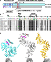
Comparisons of KRGG1 and RESC proteins. (A) Schematic diagram of full-length T. brucei KRGG1 (cyan) and RESC6 (gray) sequences from 29.13 strain with amino-terminal region RGG and RG sequences highlighted in purple and blue, respectively. The Alphafold predicted ARM/HEAT-like tandem helical repeat region is indicated by labeled brackets and the conserved ARR and LLLN regions are indicated in red and magenta, respectively. Region of the schematic covered in the multispecies sequence alignment below indicated. (B) Similarity between RESC6 and KRGG1 amino acid sequences from eight kinetoplastid species. MUSCLE alignment shows conservation in the ARR (313–315) and LLLN (332–342) regions. One hundred percent similarity is shown as black, 80%–100% as dark gray, 60%–80% as light gray and <60% as white. (C) Alphafold predicted structures from T. brucei for KRGG1 (cyan), RESC6 (gray), and RESC8 (orange). Low confidence predictions of amino- and carboxy-terminal portions of KRGG1 (including the RGG repeat domain), RESC6, and RESC8 structures are not shown.
The similarity of ARM/HEAT-like structures in RESC6 and RESC8 (McAdams et al. 2019) led us to examine Alphafold structures of other RESC components. This analysis revealed that 11 of the 21 proteins currently assigned to RESC have similar tandem helical repeats forming a right-handed superhelical structure: RESC3 (Q381A0), RESC4 (Q384R6), RESC6 (Q57ZX7), RESC8 (Q389W4), RESC9 (Q585T1), RESC10 (Q57VS6), RESC11A/11B (Q57WL2/Q57Y19), RESC12/12A (Q57Y20/Q57WL3), and RESC16 (Q585C7) (Supplemental Fig. 1). While nearly all these proteins lack identifiable motifs at the primary sequence level, the broad similarities in predicted structure suggest that these proteins are all functionally related to each other and KRGG1.
KRGG1 is essential for growth of BF T. brucei
To examine the function of KRGG1 in BF cells, we created a BF conditional null cell line that requires tetracycline (tet) for regulated expression of TAP-tagged KRGG1 from an ectopic locus. Both endogenous alleles were deleted, and a tet-regulated KRGG1-TAP allele was inserted into the ribosomal DNA (rDNA) locus. Culturing these cells in the absence of tet dramatically reduces KRGG1 expression and results in strong growth inhibition (Fig. 2A). Western analysis probing for the TAP-tag on tet-regulated KRGG1 shows strong repression after tet withdrawal, with KRGG1-TAP strongly repressed by day one and essentially undetectable by day two. These results unambiguously show that KRGG1 is essential for growth in BF cells. Glycerol gradient analysis of RECC and RESC components showed no notable changes in their abundance or sedimentation after KRGG1 repression (Supplemental Fig. 2). To confirm that the growth defect observed in these cells results from an effect on mitochondrial gene expression, we transfected a mutant gamma ATP synthase (MGA) allele into the BF KRGG1 CN background, as this circumvents the essentiality of mitochondrial gene expression in BF (Dean et al. 2013; Carnes et al. 2017). MGA expression eliminates the growth defect caused by KRGG1 repression in BF cells, with western analysis showing that KRGG1 remains repressed in -tet cells throughout the time course (Fig. 2B). These results indicate that KRGG1 function is required for normal mitochondrial gene expression.
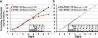
KRGG1 is essential for BF growth in vitro. (A) Growth of BF KRGG1 CN that express KRGG1-TAP in the presence of tet (black) and in which KRGG1 expression is repressed upon removal of tet (red). (Inset) Western analysis of KRGG1-TAP cell lysates from day 1 and 2, showing tet-regulation of KRGG1-TAP expression. (B) Growth of BF KRGG1 CN cells expressing mutant gamma ATP synthase allele (MGA) that circumvents the essentiality of mitochondrial gene expression. MGA KRGG1 CN cells cultured in the absence of tet (gray) grow indistinguishably from those with tet (black). (Inset) Western blot shows KRGG1 repression in −tet culture is maintained throughout the time course.
KRGG1 is required for normal RNA editing in BF T. brucei
The abundances of multiple transcripts, including edited, pre-edited, and never-edited RNAs, were assessed by RT-qPCR from multiple time points after KRGG1 repression and compared to corresponding samples with continued KRGG1 expression (Fig. 3A). KRGG1 mRNA was reduced by >99% at all time points, indicating essentially complete loss of mRNA by day one, which is consistent with western blot analyses (Fig. 2). Never-edited mRNAs ND4, COI, ND1, ND2, and ND5 remained essentially unchanged after loss of KRGG1. While most edited transcripts did not appear to change after KRGG1 repression, amplicons reporting the steady state levels of both edited ND8 and edited A6 were changed in opposite directions. Edited ND8 dramatically increased by 7.4-fold 2 d after loss of KRGG1. In contrast, edited A6 dramatically decreased fivefold 3 d after loss of KRGG1. As A6 protein is essential in BF cells (Szempruch et al. 2015), the loss of edited A6 could be responsible for the growth defect observed after KRGG1 repression. Amounts of pre-edited mRNAs were largely unchanged as has been previously observed after loss of essential RECC components (Carnes et al. 2005, 2008, 2017; Trotter et al. 2005; Salavati et al. 2006; Guo et al. 2008, 2012; Ernst et al. 2009; McDermott et al. 2015; McDermott and Stuart 2017). KRGG1 CN cells expressing MGA were also examined by RT-qPCR, as elimination of the growth defect allowed editing defects to accumulate over time (Fig. 3B). In these cells, loss of edited A6 reached 97% by day 6, indicating that KRGG1 is essential for A6 editing. An increased abundance of edited ND8 amplicon was again observed in the absence of KRGG1, peaking at 6.1-fold higher at day 2, then stabilizing at 2.4- to threefold higher on subsequent days. A 1.5 to threefold loss of MURF2 editing was detected in the KRGG1 CN MGA cell line, but not in the parental KRGG1 CN cell line, and an approximately twofold increase in ND3 editing in the parental KRGG1 CN cell line was not detected in the KRGG1 CN MGA cell line. The inconsistency of the effect of KRGG1 loss on MURF2 and ND3 editing in these cell lines suggests that the derived MGA line may have subtly diverged from parental cells during the cloning process, or that MURF2 and ND3 may not be bona fide targets of KRGG1 function. In both KRGG1 CN and derived MGA cells 12S rRNA levels were reduced approximately threefold at the end of the time course, suggesting that KRGG1 function could also play a role in mitochondrial ribosomal RNA processing and/or stabilization.
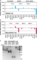
RT-PCR analyses showing the impact of KRGG1 loss on RNA editing in vivo. (A) RT-qPCR assay of BF KRGG1 CN cells showing that over 3 d repression of KRGG1 results in increases of edited ND8 amplicon and decreases of edited A6 amplicon, relative to the telomerase reverse transcriptase (TERT) mRNA internal control. Never-edited, pre-edited, and edited mRNAs are indicated by brackets. (B) RT-qPCR assay as in A but examined over 8 d in BF KRGG1 CN + MGA cells. (C) RT-PCR products corresponding to full length pre-, partially and fully edited ND8, ND3, MURF2, and A6 mRNAs from cells in which KRGG1 is expressed (+) or repressed (−). The arrowheads indicate bands where there were noticeable differences in the ND8 and A6 profiles between KRGG1 expressed versus repressed. Brackets indicate the expected size ranges from pre-edited to fully edited amplicons as noted above for each mRNA.
As RT-qPCR focuses on editing in specific regions of each transcript, we sought to examine a more complete profile of editing in KRGG1 CN cells for selected transcripts by gel electrophoresis of RT-PCR products that span the entire edited region of ND8, ND3, MURF2, and A6 (Fig. 3C). In this assay, primers anneal to never-edited regions flanking the editing domain, permitting amplification of pre-edited, partially edited, and fully edited sequences, resulting in a profile broadly reflecting multiple steps in the editing process. Consistent with RT-qPCR results, loss of KRGG1 resulted in increased ND8 editing products near the region of the expected full-length amplicon. Compared to cells expressing KRGG1, cells lacking KRGG1 accumulated more edited ND8 products in the region immediately above ∼500 bp, close to the 564 bp size expected for fully edited ND8 amplicon. Analysis of A6 editing was similarly consistent with RT-qPCR results, as loss of KRGG1 resulted in decreased A6 editing products between ∼650 to ∼750 bp, adjacent to the region of the expected full-length amplicon. Cells lacking KRGG1 had increased partially edited products around 500 bp and decreased partially edited products between 600–800 bp in comparison to cells expressing KRGG1. Relatively minor differences between KRGG1 expressed and repressed samples were observed in RT-PCR products for ND3 and MURF2, similar to their corresponding RT-qPCR results. Together, these results indicate KRGG1 function preferentially affects specific RNAs, and is essential for A6 editing. Strikingly, KRGG1 function can either impede or promote editing in different mRNA contexts.
Mutations in KRGG1 tandem helical repeat region prevent function
While the RGG domain in KRGG1 has been shown to bind RNAs, functional domains in the remainder of KRGG1 predicted to form tandem helical repeats have not been identified (Vanhamme et al. 1998). We therefore created a series of site-directed mutant versions of KRGG1 that were V5 epitope-tagged and constitutively expressed from the tubulin locus in the background of the BF KRGG1 CN cell line. Exclusive expression of the mutant allele, or wild-type (WT) control, occurs when tet is removed in these cell lines (Fig. 4; Supplemental Fig. 3). Three types of mutants were created: (i) single amino acid substitutions, (ii) multiple amino acid substitutions, or (iii) multiple amino acid deletions. We focused our mutations on either amino acids that were highly conserved in multiple kinetoplastid orthologs of KRGG1, or regions of conservation shared with RESC6. To analyze the functionality of the various KRGG1 mutants, we performed growth analysis in the presence or absence of tet. The parental CN cell line shows a severe growth defect, while expression of V5-tagged WT KRGG1 from the tubulin locus permits continuous cell growth in the absence of tet. Exclusive expression of single amino acid substitutions of highly conserved amino acids K294A, D328A, N337A, F342A, S448A, H486A, T522A, or R561A (Tb927.6.2230 numbering) resulted in weak or negligible growth defects compared to WT KRGG1. In contrast, some of the mutations in the ARM/HEAT-like region resulted in strong growth defects. Deletion of the amino acids ARR at positions 313–315 (ΔARR) that are highly conserved between KRGG1 and RESC6 resulted in a strong growth defect, as did substitution of these amino acids with RAA. Changing the last two amino acids in this triplet to alanines or aspartates, in contrast, had no appreciable growth defect. This mutation series particularly highlights A313, as it is the only residue that differs between nonfunctional RAA mutants and functional AAA mutants in this region. Similarly, deletion of 11 amino acids VTLLLNCAATF (332–342) resulted in strong growth defects when exclusively expressed, as did substitution of amino acids LLLN (334–337) with alanines. The nature of the mutation in this region is clearly important, as single substitution mutants D328A, N337A, and F342A in or near this LLLN region did not grossly alter growth when exclusively expressed. The expression level of all but one of the dysfunctional KRGG1 mutants was indistinguishable from WT, indicating that observed growth defects did not result from a reduced amount of exclusively expressed mutant KRGG1 (Supplemental Fig. 4). Because the residues required for KRGG1 function are highly conserved in RESC6, these results suggest that this region is also likely to be critical for RESC6 function. RT-qPCR from multiple time points after exclusive expression of dysfunctional KRGG1 mutants shows similar profiles to the parental BF KRGG1 CN cell line shown in Figure 3A, with the most prominent defects being decreases in edited A6 amplicon and 12S rRNA, while the increase in the ND8 edited amplicon was somewhat reduced and even observed when WT KRGG1 was exclusively expressed (Supplemental Fig. 5).
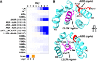
Effects of KRGG1 mutations on cell growth. (A) The Log2 ratio of cumulative growth without tet versus with tet is shown as a heat map for each day over a 7 d period. The associated scale indicates greater growth inhibition by darker shades of blue, no effect on growth by white and increased growth by darker shades or orange. Controls are the parental conditional null (CN) cell line (top row), cells exclusively expressing a wild-type (WT) KRGG1 (second row), cells with an introduction of the mutant gamma ATP synthase allele (MGA) which permits normal growth in the absence of KRGG1 (third row). Numbering for amino acid substitutions is from the Tb927.6.2230 reference sequence. (B) Location of amino acids that are critical for KRGG1 function in a portion of the Alphafold predicted ARM/HEAT-like tandem helical repeats. The top panel highlights the location of the ARR triplet, and the bottom panel highlights the LLLN region. Amino acids in these regions that are critical for function are noted. Five alpha helices in this region are labeled H1–H5 in amino- to carboxy-terminal order.
Mutations that disrupt KRGG1 function also reduce association with RESC
To assess whether the mutations that disrupt KRGG1 function alter the ability of KRGG1 to associate with RESCs, we performed immunoprecipitations (IPs) using anti-RESC13 antibody and probed for associated KRGG1 (Fig. 5). Cell stabilates of each mutant and control WT cell line were thawed and grown as before. While we noticed that expression levels of mutant KRGG1-V5 were observed to be slightly decreased in comparison to WT, we confirmed that growth of these cells with or without tet was as previously described. In this assay, WT KRGG1-V5 was readily detected in anti-RESC13 IPs, as was the functional AAA (313–315) mutant (Fig. 5A). In contrast, mutants that render KRGG1 nonfunctional (ΔARR, ARR → RAA, ΔVTLLLNCAATF, and LLLN → AAAA) all had broadly reduced association with RESC. Nonfunctional KRGG1 mutants were only detected in anti-RESC13 IPs after long exposures, indicating reduced association with RESCs. In a replicate experiment, nonfunctional KRGG1 mutants were not detected even in long exposures, indicating that very little of these mutant proteins associate with RESCs (data not shown). Parental KRGG1 CN cell line lacks a V5-tagged allele and served as a negative control in these experiments. Because the nonfunctional KRGG1 mutant proteins also have reduced abundance in cleared lysates relative to WT, we quantified the reduction in KRGG1 association in anti-RESC13 IPs by first normalizing to the amounts in input cleared lysates, then comparing relative to WT KRGG1 sample (Fig. 5B). While the amount of KRGG1 associated with RESC was reduced to <25% of wild-type amount for all the dysfunctional mutants, this overestimates the amount of nonfunctional KRGG1 mutant association with RESC because KRGG1 WT signal is saturated in long exposures in which nonfunctional KRGG1 mutants can be detected. The amount of dysfunctional KRGG1 mutant protein that is associated with RESC is less than 10% of the wild-type amount when the KRGG1 signal in anti-RESC13 IPs is normalized to the RESC13 IP signal. Thus, mutations that disrupt KRGG1 function also reduce association with RESCs. Probing with either anti-RESC1 or anti-RESC13 antibodies showed that anti-RESC13 IPs successfully isolated RESCs. To quantify the abundances of RESC1 and RESC13 in anti-RESC13 IPs, they were both normalized to the amount of control HSP70 observed in input cleared lysates, then compared relative to WT KRGG1 sample. These results indicate that RESCs were effectively isolated in all samples, in contrast to the reduced signals observed for nonfunctional KRGG1 mutants in anti-RESC13 IPs.
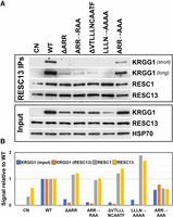
Effects of KRGG1 mutations on RESC association. Anti-RESC13 immunoprecipitations (IPs) co-isolate KRGG1 that is detected via a carboxy-terminal V5-epitope tag. (A) Western analyses of anti-RESC13 IPs (top panel) probed for KRGG1 show stronger signal for WT and functional ARR → AAA mutants relative to dysfunctional mutants in both short and long exposures; probing for either RESC1 or RESC13 shows the isolation of RESCs. Western analyses of cleared lysate inputs used for IPs (bottom panel) probed for the amounts of KRGG1, RESC13, or mitochondrial HSP70 control present in each sample. (B) Densitometric quantitation of the above western blots, with KRGG1 signal in IP (long exposure) normalized to either KRGG1 signal in input or RESC13 signal in IP, RESC1, and RESC13 signal in IP normalized to HSP70 signal in input, and all reported relative to KRGG1 WT sample.
DISCUSSION
We report here that KRGG1 is essential for growth and editing in vivo in T. brucei, and its function has differential effects on various mitochondrial RNAs. Rescue from the growth defect due to the loss of KRGG1 by MGA expression shows that KRGG1 is essential for editing and mitochondrial gene expression. The observed loss of 12S rRNA following the repression or mutation of KRGG1 suggests that its function not only impacts mRNA editing but may also impact mitochondrial translation. However, our results resolve the uncertainty about whether KRGG1 functions in RNA editing that stemmed from limitations of RNAi knockdowns in previous T. brucei experiments. Use of MGA expression also permits more detailed examination of RNA editing defects after KRGG1 loss, thus revealing increased abundance of edited ND8 amplicon and a dramatically decreased abundance of edited A6 amplicon. The strong differential effects on A6 and ND8 editing after KRGG1 repression highlights both the complexity of the RNA editing process and the specialized role that KRGG1 plays within it. The revelation that KRGG1 shares sequence similarity to RESC6 has been extended by Alphafold structural analysis, which shows that a right-handed superhelix of tandem helical repeats is shared by multiple RESC components. The predicted nucleic acid-binding capabilities of the KRGG1 helical repeat structure, combined with the previously demonstrated RNA-binding ability of the RGG domain provide critical clues for the function of in RNA editing of specific mRNAs. Furthermore, mutational analysis of the tandem helical repeat region shows that this newly identified domain is essential for KRGG1 function and association with RESCs and by extension the similar region in RESC6 appears likely to be essential for editing.
Alphafold structures provide context for the mutations that disrupt KRGG1 function. Disruptive mutations to either the ARR triplet or LLLN region are located in α-helices at the beginning of the tandem helical repeat region of KRGG1 that lies carboxy-terminal to the RGG repeats, with ARR in α-helix H2, and the LLLN region in the α-helix H3 (Fig. 4). Mutations in the beginning of the repeat structure could disorder the packing of helices onto each other, grossly perturbing the overall structure, or they could disrupt the junction between the RGG repeat domain and the tandem helical repeat domain. Our mutational analysis of the ARR region identified the initial A313 as critical for KRGG1 function, as replacement of this alanine with a larger arginine in the RAA mutant resulted in a strong growth defect, while the AAA mutant permitted normal growth when exclusively expressed. In the context of the predicted KRGG1 structure, the change from smaller A313 to larger R313 might sterically disrupt the packing of α-helix H2 with H1 and/or H3. Despite the broad conservation of R314 and R315, replacement of both residues with alanines did not prevent KRGG1 function, and the predicted structure suggests a possible reason why. One side of the α-helix H2 contains seven positively charged arginines/lysines, of which R314 and R315 are only two. Thus, the remaining positive charged K304, R307, R311, R318, and R319 likely mask the loss of R314 and R315. The deletion of ARR prevents normal KRGG1 function, and likely disrupts interactions between helices H1–H3 of the tandem helical repeat region by placing amino acids out of register within this structural context. Deletion of the LLLN region (ΔVTLLLNCAATF) essentially eliminates helix H3, which is centrally located between H1, H2, H4, and H5, interacting with all of them. The disruptive effects of the LLLN to AAAA mutation at positions 334–337 appear likely to prevent the hydrophobic interactions of L334 with helix H1 and of L335 and L336 with helix H4. The loss of these hydrophobic interactions could then prevent proper alignment or packing within the tandem helical repeats.
Although the amino-terminal RGG domain of KRGG1 is an entirely low-confidence unstructured region in Alphafold prediction (Supplemental Fig. 1), studies in other RGG-containing proteins provide context for how these repeats may function in KRGG1 and suggest a mechanism for the observed effects on A6 and ND8 editing. Solution structure of the complex between the FMRP RGG domain and G-quadruplex-containing RNA has been solved, revealing that RGG is positioned along the major groove of the RNA duplex adjacent to G-quadruplex (Phan et al. 2011). G-quadruplexes are arrangements of four guanines that are formed through cyclic Hoogsteen hydrogen-bonding interactions, and the planar G-quadruplexes can stack on top of one another to forming four-stranded helical structures (Rhodes and Lipps 2015). While it remains unclear how generalizable this interaction is, a high preponderance of G-quadruplex sequences has been noted in pre-edited mRNAs that are resolved by the process of editing (Leeder et al. 2016). Both A6 and ND8 pre-edited mRNAs have such G-quadruplex sequences that have been experimentally characterized (Leeder et al. 2016), and the sizes of RT-PCR products that have altered editing after loss of KRGG1 in Figure 3C are consistent with regions in these mRNAs proximate to G-quadruplexes (Supplemental Fig. 6). Additional experiments will be needed to determine whether direct interactions between these RNA editing substrates and KRGG1 exist in vivo. A combination of both the RGG and ARM/HEAT-like domains could provide binding specificity for A6 and ND8 that mediates the action KRGG1 has on these mRNAs.
The results from this work clearly establish a role for KRGG1 in the RNA editing process, and mutations that disrupt function also disrupt association with RESCs. However, the nature and dynamics of its interaction with RESCs are unclear, as exemplified by the lack of detecting KRGG1 interactions with other RESC components in yeast two-hybrid experiments and the reduced association of KRGG1 with isolated RESC complexes upon RNase digestion (Hashimi et al. 2008; Ammerman et al. 2012; Aphasizheva et al. 2014). RNase digestion also affects the association of known RESC components, resulting in reduced interactions in vitro in the absence of RNA (Aphasizheva et al. 2014) which may imply dynamic and diverse associations among RESC proteins. Furthermore, the stoichiometric ratios of RESC components vary widely, suggesting that RESC composition is not monolithic and is likely dynamic (Dubey et al. 2021). Because RNAs themselves can be a critical element for facilitating the formation of ribonucleoprotein complexes, the additional data presented here suggests that KRGG1 may be a RESC component with a dynamic association profile (Garcia-Jove Navarro et al. 2019). Overall, KRGG1 function is clearly intertwined with those of RESC and RECC since all functionally impact the editing of substrate RNAs. Mutations that prevent KRGG1 function affect editing catalyzed by RECCs and greatly reduce KRGG1 association with RESCs, suggesting that KRGG1's function involves direct interaction with RESCs. Additional experiments are needed to address the physical and functional interactions between KRGG1, RESCs and RECCs.
The data presented here provide new insights into possible modes of action of how KRGG1 might function. Our mutation data indicate that the ARM/HEAT-like domain in KRGG1 is essential thus identifying a domain that likely binds RNA in a manner distinct from the amino-terminal RGG domain. These two domains together could enhance specificity of RNA binding, perhaps including preferential binding of editing substrates like A6 or ND8. The broad conservation of right-handed superhelical tandem helical repeat structures among multiple RESC proteins suggests that these proteins are paralogs that have specialized but related functions. These structures are reminiscent of PPR proteins that are widespread in mitochondria and chloroplasts of plants and have been characterized as sequence-specific RNA-binding proteins involved in multiple aspects of RNA metabolism, including C to U RNA editing (Kotera et al. 2005; Fujii and Small 2011; Wang et al. 2021). PPR proteins also form right-handed superhelical spirals of tandem α-helical repeats, with individual α-helical modules specifically recognizing a single nucleotide of a target RNA, and multiple modules combining to recognize a specific RNA sequence (Yin et al. 2013). These structural similarities suggest the possibility that KRGG1 and related RESC proteins could recognize specific RNA sequences to facilitate their editing. This hypothesis fits the specific effects observed on editing of A6 and ND8 mRNAs after KRGG1 repression. While the precise nature of how KRGG1 functions will require additional experiments to be elucidated, the results presented here indicate that understanding the details of how KRGG1 binds RNA will likely shed light on the function of multiple related RESC proteins.
MATERIALS AND METHODS
Sequence and structure analyses
Amino acid sequences for RESC6 and KRGG1 from multiple kinetoplastid species were obtained from TriTrypDB and analyzed using Geneious Prime (see Supplemental Table 2 for GeneIDs). MUSCLE alignment was highlighted with Blosum 62 matrix, threshold 1. Alphafold structures were taken from AlphaFold DB version 2022-06-01, created with the AlphaFold Monomer v2.0 pipeline (Jumper et al. 2021; Varadi et al. 2022). Low and very low confidence residues (pLDDT scores below 70) at amino- and carboxy-termini were removed for clarity in Figure 1 structures displayed using ChimeraX (Pettersen et al. 2021).
Generation and growth of transgenic cell lines
The procedure for generating a conditional KRGG1 null cell line follows our previously published approach (Merritt and Stuart 2013). Primers used to make constructs for transgenic cell lines are in Supplemental Table 3. Briefly, constructs to knockout KRGG1 alleles were generated with KRGG1 5′ and 3′ UTRs flanking either hygromycin (HYG) or blasticidin (BSD) resistance genes in fusion PCR reactions. WT KRGG1 ORF was PCR amplified from 29.13 genomic DNA and cloned into pLEW100V5(BLE)GW-CtermTAP that adds a tandem affinity purification (TAP) tag on the carboxyl terminus of the KRGG1 ORF and allows for tet-regulatable expression from the rRNA locus (McDermott et al. 2015). Sequencing of the KRGG1 ORF cloned from 29.13 revealed an internal deletion of four RGG repeats in the amino-terminal RGG repeat region of amino acids 170–221 (when compared to the Tb927.6.2230 sequence); this alternate KRGG1 sequence is also present in the published pLew79-KRGG1-MHTAP plasmid independently cloned from 29.13 (Hashimi et al. 2008). Sequential transfections into the BF SM427 background generated BF KRGG1 CN cell line: the first KRGG1 allele was knocked out using the HYG construct, then the NotI-linearized pLEW100v5(BLE)-KRGG1-TAP plasmid introduced tet-regulatable ectopic KRGG1-TAP (selected using phleomycin), and finally the second KRGG1 allele was knocked out using the BSD construct. To generate exclusive expression cell lines, WT KRGG1 lacking the stop codon and flanked in frame with attB Gateway recombination sites, was PCR amplified and transferred into pDONR221. pENTR-KRGG1 was used in LR reactions with the destination vectors pHD1344tub(PAC)GW-Cterm3V5, which allow for constitutive expression of carboxy-terminal 3xV5 tagged proteins in the β-tubulin locus (McDermott et al. 2015). The resulting pHD1344tub(PAC)-KRGG1-Cterm3V5 plasmid was then used as a template for site-directed mutagenesis (QuikChange II Kit; Agilent). NotI-digested plasmids were then transfected into the BF KRGG1 CN cell line and selected by puromycin resistance. Constitutive expression of KRGG1-3xV5 protein (either WT or mutant) was confirmed by western blot. Drug concentrations used were 2.5 µg/mL G418, 2.5 µg/mL hygromycin, 2.5 µg/mL phleomycin, 1.25 µg/mL blasticidin, 0.5 µg/mL tetracycline, and 0.1 µg/mL puromycin. BF cells were grown in HMI-9 (Hirumi and Hirumi 1989) with 10% FBS at 37°C, 5% CO2. For growth curve analysis, cell density was measured using a Coulter Counter. BF were reseeded at 2 × 10⁵ cells/mL in 5 mL every day. To generate cell growth heat maps, cumulative growth numbers for −tet and +tet cultures were calculated for each time point. Next, log2 of the ratio of −tet to +tet was calculated, and this number was converted to blue to orange scale using conditional formatting in Microsoft Excel.
SDS-PAGE and western blotting
SDS-PAGE loading buffer was added to cleared whole cell lysates, or to samples containing purified protein complexes and resolved on 10% SDS-PAGE gels (Criterion Tris-HCl, Bio-Rad). For Western analysis, resolved proteins were transferred to Immobilon-P PVDF membranes (Millipore), and probed using antibodies described in Supplemental Table 4. Blots were developed with an Enhanced Chemiluminescence Kit (Thermo Scientific) per the manufacturer's instructions and imaged using x-ray film (McKesson).
RNA isolation, RT-qPCR, and RT-PCR analyses
Total RNA was harvested using TRIzol and treated with TURBO DNase (Life Technologies) according to manufacturer's instructions. RNA integrity was confirmed using an RNA nanochip on a BioAnalyzer (Agilent Technologies). An amount of 2 µg of total RNA was reverse-transcribed using TaqMan Reverse Transcription Reagents and MultiScribe Reverse Transcriptase (Life Technologies), preamplified in multiplex Specific Target Amplification (STA) reactions using TaqMan PreAmp Master Mix (Life Technologies) and treated with Exonuclease I (NEB). The abundance of reference, never-edited, pre-edited and edited transcript cDNAs were then analyzed by high-throughput real-time PCR on the BioMark HD system as previously described (McDermott et al. 2015). Calculations of RNA levels in samples following tet withdrawal relative to the presence of tet, were done using the 2 [−ΔΔC(T)] method (Livak and Schmittgen 2001) using TERT as an internal reference. Technical replicates (at least two) of each cDNA sample were assayed for each target and internal reference per experiment and C(T) data averaged before performing the 2 [−ΔΔC(T)] calculation. Additionally, experiments were repeated using at least two biological replicates. For RT-PCR reactions in Figure 3C, 1 µg of total RNA (DNase treated as above) was converted to cDNA with gene-specific reverse primer and MultiScribe Reverse Transcriptase followed by 35 cycles of PCR amplification and analysis of products on 2.75% agarose TBE gels stained with ethidium bromide. Cycling conditions were tested to ensure that 35 cycles of amplification did not introduce artifacts. Gels were visualized using AlphaImager EP (AlphaInnotech). Primers for all above experiments are described in Supplemental Table 3.
Immunoprecipitations and western analyses
For each immunoprecipitation, cells were harvested after growth without tet for 48 h and flash frozen on liquid nitrogen and stored at −80°C. Cleared lysate was prepared using 2.4 × 10⁸ cells in 1 mL IPP150 (10 mM Tris-HCl pH 8.0, 150 mM NaCl, 0.1% Nonidet P-40, Roche Complete protease inhibitors) with 6.25 µL RNasin (Promega N261A), 1 mM DTT, and 1% Triton X-100. Lysates were cleared by centrifugation at 12,000 rpm at 4°C. An amount of 50 µL of each cleared lysate was added to 50 µL 2× SDS sample buffer for control input samples, with 10 µL loaded onto 10% Criterion gels for western analysis. The remaining cleared lysate was incubated for 2.5 h with anti-RESC13 magnetic beads. To prepare magnetic beads for each anti-RESC13 immunoprecipitation, 10 µL of Protein G Mag Sepharose Xtra (GE Healthcare) was washed three times with 100 µL of IPP150 then resuspended in 25 µL IPP150. Next, 1 µL anti-RESC13 rabbit polyclonal antibody was added to washed Protein G Mag beads and allowed to incubate overnight at 4°C with mixing. After overnight antibody binding, beads were washed four times with 1 mL IPP150. After incubation of cleared lysate with anti-RESC13 magnetic beads, supernatant was removed, and beads were washed four times with 1 mL IPP150. Complexes bound to beads were eluted with 25 µL of 2 × SDS sample buffer, heated for 5 min at 95°C, with half of that volume loaded onto 10% Criterion gels for western analysis.
SUPPLEMENTAL MATERIAL
Supplemental material is available for this article.
ACKNOWLEDGMENTS
The authors would like to thank Madison Meuler for assistance with western blots, H. Ulrich Göringer for sharing G-quartet structure images of A6 and ND8, and Laurie Read for sharing antibodies against RESC11A and RESC13. This work was supported by National Institutes of Health grant R01AI014102 to K.S. and Seattle Children's Research Institute.
Footnotes
-
Article is online at http://www.rnajournal.org/cgi/doi/10.1261/rna.079418.122.
-
Freely available online through the RNA Open Access option.
- Received August 15, 2022.
- Accepted November 11, 2022.
This article, published in RNA, is available undera Creative Commons License (Attribution-NonCommercial 4.0 International), as described at http://creativecommons.org/licenses/by-nc/4.0/.
REFERENCES
MEET THE FIRST AUTHOR

Meet the First Author(s) is an editorial feature within RNA, in which the first author(s) of research-based papers in each issue have the opportunity to introduce themselves and their work to readers of RNA and the RNA research community. Jason Carnes is the first author of this paper, “KRGG1 function in RNA editing in Trypanosoma brucei.” Jason did his PhD in the labs of Leslie Leinwand and Michael Yarus at the University of Colorado, Boulder, investigating the use of RNA aptamers to inhibit translation termination, and did a brief post-doc with Rob Knight at UC Boulder examining the evolutionary relationships between biological sequences with a focus on tRNA synthetases. Jason then moved to Seattle to work for Ken Stuart, where he continues to work at Seattle Children's Research Institute investigating several aspects of RNA editing in trypanosomes.
What are the major results described in your paper and how do they impact this branch of the field?
Our paper shows that KRGG1 is required for normal RNA editing in Trypanosoma brucei, and that it is a distant homolog of RESC6, which is one of several proteins in RNA editing substrate complexes. We identify that the region of KRGG1 most similar to RESC6 is critical to sustain its association with RESCs, RNA editing, and cell growth. This region of similarity is predicted to form an ARM/HEAT-like superhelical structure that we also discovered in many other RESC proteins. Together, these results resolve uncertainty in the field regarding the role of KRGG1 in RNA editing and indicate that the function of structurally similar RESC proteins may also require the newly identified ARM/HEAT-like region that is required for KRGG1 function.
What led you to study RNA or this aspect of RNA science?
When I started my PhD studies at the University of Colorado, Boulder, I was primarily interested in human gene therapy as a field of study. My thesis project was to develop RNA aptamers to target translation termination as a potential therapy to correct nonsense mutations. The more I learned about RNA biology, the more my career path bent toward it. I was a cochair of the RNA Club at UC Boulder as a grad student, and exposed to the work in the labs of Olke Uhlenbeck, Norm Pace, and Tom Cech among others. After I graduated, I moved to Seattle to work with Ken Stuart on RNA editing and have remained focused on understanding this amazingly complex system ever since.
During the course of these experiments, were there any surprising results or particular difficulties that altered your thinking and subsequent focus?
We were initially surprised to identify the homology between KRGG1 and RESC6, which turned our attention on to other RESC proteins that have similar predicted structures. Finding mutations in KRGG1 that resulted in functional consequences was a particular challenge, as most of that work was done before the Alphafold predicted structures were available.
What are some of the landmark moments that provoked your interest in science or your development as a scientist?
I cannot recall a time when I wasn't interested in the biological world and understanding how it works, probably in part because my mother was a middle school life sciences teacher and she fostered my curiosity. I distinctly remember when I decided to focus on science as a career, though, during my sophomore year in high school. My life sciences biology course was taught by Rodney Epp, and he had an incredible ability to make science exciting and fun. Great teachers make a difference.
If you were able to give one piece of advice to your younger self, what would that be?
Every experiment involves opportunity cost. Be prepared to change your experimental priorities if you identify a better avenue for exploration before the current work is finished.
Are there specific individuals or groups who have influenced your philosophy or approach to science?
So many people have informed the way I approach science, that I find it difficult to summarize succinctly. Michael Yarus said simply that you need a positive control, a negative control, and an experiment. Bob Thompson really helped me see the proper way to break down an experiment to troubleshoot problems. Ken Stuart keeps me thinking about the bigger picture and the value of different perspectives to gain insight. Massimo Buvoli, Steve Langer, and Leslie Leinwand taught me how to keep things fun in the lab and stay positive in the face of inevitable challenges.
What are your subsequent near or long-term career plans?
I plan to continue my research in RNA editing for as long as they'll let me do it.




