


ADARs mediate distinct RNA editing activity and gene regulation in the Caenorhabditis elegans germline
- 1Genome, Cell and Developmental Biology Graduate Program, Indiana University, Bloomington, Indiana 47405, USA
- 2Department of Biology, Indiana University, Bloomington, Indiana 47405, USA
- Corresponding author: hahundle{at}iu.edu
-
Handling editor: Eric Phizicky
Abstract
Tissues rely on unique landscapes of gene regulation to allow the organism to correctly develop, function, and respond to changes. One component of these gene regulatory networks is RNA binding proteins (RBPs), which bind and modify RNA molecules leading to changes in the cellular fate of transcripts. The adenosine deaminase acting on RNA (ADAR) family of RBPs modify RNAs by catalyzing the deamination of adenosine (A) to inosine (I), known as A-to-I RNA editing. Prompted by recent evidence that ADARs play important roles in germline biology, we profiled editing activity of the A-to-I editing enzyme ADR-2 on transcripts in the Caenorhabditis elegans germline. These analyses revealed that many germline editing events are distinct from editing events in other tissues; however, the previously described role of the inactive deaminase ADR-1 in regulating editing activity by ADR-2 is conserved in the germline. We find that complete loss or misregulation of editing has little effect on the expression of edited transcripts within the germline; however, loss of ADARs results in the misexpression of several unedited germline transcripts. Intriguingly, further investigation suggests that these expression changes are buffered at the translational level. In all, the results of this study suggest that ADARs show unique activity in the C. elegans germline and that compensatory mechanisms exist to lessen the immediate consequences of loss of ADAR function within the germline.
Keywords
INTRODUCTION
RNA-level regulation of gene expression is essential in coordinating proper development and organismal function. In particular, RNA regulation is important in tissue diversification (Franks et al. 2017). One mechanism of RNA regulation important for proper tissue development is RNA modifications, by which RNAs are chemically altered to affect their cellular fate (Porat 2025).
Adenosine-to-inosine (A-to-I) RNA editing is one of the most abundant RNA modifications in metazoans (Zhang et al. 2023) and is catalyzed by the adenosine deaminase acting on RNA (ADAR) family of RNA binding proteins (Datta et al. 2023). A-to-I editing can have various effects on transcript fate (Eisenberg and Levanon 2018). As inosine is read as guanosine by the translational machinery, editing in coding regions of transcripts can lead to changes in codons, resulting in changes in the amino acid sequence of the encoded peptide (Lewis 2023). While this “recoding” editing can have important impacts, A-to-I editing is abundant within noncoding regions of transcripts (Bazak et al. 2014). Noncoding editing primarily affects RNA processing and regulation and can lead to changes in gene expression, which ultimately result in physiological changes at the cell, tissue, and organismal levels (Rueter et al. 1999; Yang et al. 2006, 2017; Lev-Maor et al. 2007; Scadden 2007; Hsiao et al. 2018; Liu et al. 2024). A-to-I editing patterns and frequency are known to differ throughout developmental stages and tissues (Lev-Maor et al. 2007; Ekdahl et al. 2012; Tan et al. 2017; Rajendren et al. 2021). As such, tissue- and cell type–specific studies have proven useful in identifying novel molecular and biological consequences of editing (Fei et al. 2016; Deffit et al. 2017; Chen et al. 2024).
In addition to A-to-I editing, ADARs are double-stranded RNA (dsRNA) binding proteins and can target dsRNAs ranging from small RNAs and their precursors to longer transcripts containing intramolecular duplexes (Marônek et al. 2025). dsRNA binding by ADARs can impact transcripts by competing with other RNA binding factors for targets or by recruiting other RNA binding factors to targets (Karlström et al. 2024; Mahapatra et al. 2025). These interactions can ultimately lead to changes in transcript processing and stability. As such, several editing-independent effects of ADARs have also been shown to play important roles in tissue development and functions (Capshew et al. 2012; Cho et al. 2018; Deng et al. 2020; Erdmann et al. 2024).
Due to the severe neural defects originally reported upon loss of ADARs in mammals (Burns et al. 1997; Higuchi et al. 2000), tissue-specific studies have mainly focused on the nervous system (Liscovitch et al. 2014; Deffit et al. 2017; Sapiro et al. 2019; Rajendren et al. 2021). However, transcriptomic studies have identified ADAR expression and editing events across a wide range of human tissues (Tan et al. 2017). Recently, emerging evidence from mammalian studies has pointed to biological roles for ADARs in germline development and function (Snyder et al. 2020; Nelson et al. 2022). While previous studies have identified editing events in germline tissues, the molecular consequences of ADARs on germline transcripts remain largely unknown.
The nematode Caenorhabditis elegans is an ideal model system for assessing the impacts of ADARs as null mutants are viable (Tonkin et al. 2002). While A-to-I editing and ADAR function have been studied extensively at the whole animal level in C. elegans (Washburn et al. 2014; Whipple et al. 2015; Zhao et al. 2015; Rajendren et al. 2018; Ganem et al. 2019; Eliad et al. 2024; Schiksnis et al. 2024), and in neural tissues (Deffit et al. 2017; Rajendren et al. 2021), there is little understanding of ADAR activity in the germline. Given emerging evidence that ADR-2, the sole A-to-I editing enzyme in C. elegans, is expressed widely in germline tissues (Eliad et al. 2024) and plays roles in regulating germline processes (Reich et al. 2018; Fischer and Ruvkun 2020; Erdmann et al. 2024), this study aimed to investigate how ADR-2 regulates transcripts in the C. elegans germline. Using an unbiased, high-throughput approach, we profile the molecular targets of germline ADARs and characterize the consequences of ADAR function on the expression and translation of these transcripts.
RESULTS AND DISCUSSION
ADARs are expressed throughout the C. elegans adult germline
In a previous study, adr-2 mRNA expression was found to be significantly reduced in young adult neural cells compared to L1 neural cells; however, the total adr-2 mRNA expression in the animal remained unchanged (Rajendren et al. 2021), begging the question of where most adr-2 expression is taking place in young adult animals. As the onset of young adulthood corresponds to reproductive maturity in C. elegans (Pazdernik and Schedl 2013), we were curious whether this expression could be coming from the germline. To determine the contribution of the germline to total ADAR expression in young adult animals, mRNA expression of the active deaminase adr-2 and the other C. elegans ADAR family member, the inactive deaminase adr-1 (Washburn et al. 2014), was measured in wild-type animals and glp-4(bn2) temperature-sensitive mutant animals which fail to develop germ cells when raised at a restrictive temperature (25°C) (Beanan and Strome 1992). Genomic deletion mutants of adr-1 and adr-2 were included as negative controls. Strikingly, germline-lacking glp-4(bn2) animals show a significant decrease in adr-2 expression compared to germline-containing wild-type animals (Fig. 1A, left), suggesting that the majority of adr-2 mRNA expression in young adult animals is within the germline. Similar results were seen for adr-1, with glp-4(bn2) germline-lacking animals showing similar adr-1 mRNA levels to those detected in adr-1(-) animals, a significant decrease compared to wild-type, germline-containing animals (Fig. 1A, right). Together, these data suggest that ADARs are abundantly expressed at the mRNA level in the germline of young adult animals. However, as mRNA expression does not always scale with protein expression, and as many germline transcripts are translationally repressed and stored for use in the early embryo (Quarato et al. 2021), it is important to confirm whether ADARs are also expressed at the protein level in the germline.
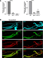
ADARs are abundantly expressed in the young adult C. elegans germline. (A) Relative mRNA expression of adr-2 (left) and adr-1 (right) in wild-type, germline-lacking glp-4 temperature-sensitive mutant, and adr null mutant young adult animals raised at the restrictive temperature of 25°C. adr expression is normalized to the housekeeping gene gpd-3. Data represent three biological replicates. Significance determined via one-way ANOVA, (****) P ≤ 0.0001. (B) Representative confocal images of young adult (left) and L4 (right) extruded germlines stained for DAPI (top, cyan), V5::ADR-1 (top middle, green), and 3xFLAG::ADR-2 (bottom middle, red). (Bottom panel) Signals of ADR-1 and ADR-2 panels merged. Dotted lines denote proximal germline region containing oocytes (young adult) or sperm (L4). Each image includes two gonad arms and the intestine from the same animal. Scale bars, 50 μm.
Recently, we have shown that ADR-2 protein is expressed in germ nuclei throughout gametogenesis, and in mature oocytes but not in mature sperm (Erdmann et al. 2024). To determine where ADR-1 is expressed in the germline and whether it correlates with ADR-2 expression, germlines were extruded from young adult and L4 animals, and immunostaining was performed for both V5-tagged ADR-1 and 3xFLAG-tagged ADR-2. ADR-1 shows a similar expression pattern to that of ADR-2, with expression in germ nuclei throughout gametogenesis and in oocytes (Fig. 1B, left) but not in sperm (Fig. 1B, right). While both ADARs show a strong nuclear signal, ADR-1 also appears to be distributed throughout the cytoplasm (Fig. 1B) while, consistent with previous reports, ADR-2 appears more restricted to the nucleus (Eliad et al. 2024; Erdmann et al. 2024).
Profiling A-to-I RNA editing activity on germline transcripts
Given our results that both ADR-2 and ADR-1 are expressed throughout the germline, we sought to determine whether ADARs are active in catalyzing and regulating A-to-I RNA editing in the germline. To profile A-to-I editing in the young adult C. elegans germline, germline RNA sequencing was performed. Sequencing libraries were prepared from dissected germline samples from wild-type and adr-2(-) animals, and sequencing reads were aligned to a reference genome. To identify editing sites in germline transcripts, we used SAILOR, a bioinformatic program that identifies high-confidence A-to-I editing sites (Deffit et al. 2017; Kofman et al. 2023). Putative A-to-I editing sites were restricted to sites with a reproducible confidence level ≥75% from SAILOR in all three biological replicates (1098 putative editing sites) of the wild-type germline samples. As adr-2(-) animals lack A-to-I editing (Tonkin et al. 2002; Washburn et al. 2014), the adr-2(-) RNA-seq data serve as a control for the computational analysis. Consistent with this, SAILOR analysis on the RNA-seq from adr-2(-) germlines identified 86 sites that were also identified in the wild-type animals, which were in turn removed as false positives. These analyses identified a final list of 1012 high-confidence A-to-I RNA editing sites in 71 germline transcripts (Supplemental Table S1). Similar to editing site analysis from adult whole worms (Schiksnis et al. 2024) and adult neural cells (Rajendren et al. 2021), the majority of germline editing sites were located in noncoding regions of transcripts, primarily the 3′ untranslated region (UTR) (Fig. 2A; Supplemental Table S1).
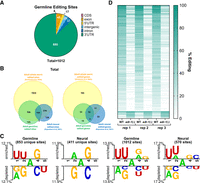
ADR-2 edits distinct targets in the germline and is regulated by ADR-1. (A) Genic distribution of high-confidence germline editing sites identified in RNA-seq data from germlines of wild-type animals. (B) Comparison of edited sites (left) and edited genes (right) between adult germline samples (this study, green), adult neural samples (Rajendren et al. 2021) (blue), and adult whole worm samples (Schiksnis et al. 2024) (yellow). (C) Two Sample Logo analyses comparing flanking nucleotides of germline and/or neural editing sites to flanking nucleotides of nearby random adenosines. Favored (above) and disfavored (below) nucleotides are shown on the x-axis. Letter height corresponds to the difference between the positive and background inputs. Statistical significance was determined by a two-sample t-test, P < 0.05, with no Bonferroni correction. (D) ADR-1-regulated germline editing sites. Percent editing at individual germline editing sites for three independent biological replicates of RNA-seq data from wild-type and adr-1(-) germlines.
Comparing our list of germline editing sites to a previously published data set from adult worms (Schiksnis et al. 2024), we found a strong overlap in edited sites and edited genes, as would be expected because the adult samples were from the entire animal, and thus, include the germline (Fig. 2B; Supplemental Table S1). To get a better idea of how germline editing compares to other tissues, we also compared our list of germline editing sites to a previously published data set from adult C. elegans neural cells (Rajendren et al. 2021). While there was some overlap between the neural and germline editomes (159 sites in 22 genes), the majority of neural and germline editing sites were mutually exclusive, with 411 sites and 44 genes exclusively identified in the neural editome and 853 sites and 49 genes exclusively identified in the germline editome (Fig. 2B; Supplemental Table S1). One possibility for the identification of unique germline-edited genes is that the genes are exclusively expressed in the germline and thus unable to be edited in other tissues. To determine whether tissue-specific expression was driving identification of germline-specific editing, we assessed the expression patterns of the 49 germline-specific edited genes and found that all of these genes are also expressed in neurons in young adult animals (Supplemental Table S1; Ghaddar et al. 2023). These data demonstrate that while some A-to-I editing sites and edited genes are conserved across tissues, there are also tissue-specific differences in editing patterns, which may allow for unique biological consequences of editing in different tissues. Consistent with these tissue-specific differences, examining the nucleotides immediately adjacent to the editing sites revealed differences in the favored and disfavored nucleotides surrounding editing sites unique to each tissue (Fig. 2C). Interestingly, these differences were not observed by simply analyzing nearest neighbors of the complete list of editing sites in each tissue, presumably due to similarities in the editing sites shared between the tissues (Fig. 2C). This work suggests that there may be factors important for specific regulation of ADAR activity in a given tissue.
ADR-1 has been shown to regulate A-to-I editing by ADR-2 in neural tissues (Washburn and Hundley 2016; Rajendren et al. 2021). Due to the differences in ADR-2 editing patterns seen between neural and germline tissues, we wanted to determine whether ADR-1 regulates editing of germline transcripts. We assessed editing levels for germline-edited sites in wild-type and adr-1(-) germlines by performing variant calling on germline RNA-seq data. Variants mapping to our 1012 high-confidence editing sites were selected for further analysis. To ensure identification of only robust changes in editing, regulated sites were defined as those with read coverage of >10 reads in both wild-type and adr-1(-) sequencing data and exhibiting a greater than |5%| difference in editing between wild type and adr-1(-) for all three biological replicates. Upon applying these parameters, 205 germline editing sites were designated as ADR-1-regulated (Fig. 2D; Supplemental Table S2). ADR-1-regulated sites were found within 36 of the 71 germline-edited genes. For the vast majority of these sites (192/205), editing decreased upon loss of adr-1, suggesting that ADR-1 promotes ADR-2 activity at these sites in wild-type germlines. The remaining 13 sites showed an increase in percent variant reads in adr-1(-) germlines compared to wild type, suggesting ADR-1 inhibits editing at these sites in wild-type germlines. However, it is important to note that while these data indicate that ADR-1 regulates A-to-I editing by ADR-2 in the germline, the ADR-1-regulated editing sites in the germline both overlap with editing sites shared between the germline and neurons and ones that are specific to the germline, suggesting that ADR-1 is likely not a factor that could be contributing to editing site specificity (Fig. 2C) in a given tissue.
Together, these data demonstrate that A-to-I editing has context-dependent characteristics, such as substrate sequence, that vary by tissue, but also RNA binding protein regulators that function across tissues. To understand the germline- and neural-specific functions and deposition of RNA editing sites, future experiments should seek to identify specific regulators of editing in these tissues.
ADR-2 regulates expression of ribosomal RNA
To investigate whether A-to-I editing can affect mRNA expression of germline transcripts, we performed differential gene expression analysis between wild-type and adr-2(-) germlines. Edited genes show minimal changes in expression between wildtype and adr-2(-) (Fig. 3A; Supplemental Table S3), with only one edited gene (gid-7) showing a significant change in expression (Padj ≤ 0.05, log2 fold change ≥0.5). This suggests that A-to-I editing does not directly impact germline mRNA expression of most edited transcripts in C. elegans.
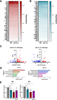
ADARs regulate germline gene expression. (A,B) Expression of edited genes in adr-2(-) germlines compared to control (WT) germlines (A) and adr-1(-) germlines compared to control (WT) germlines (B). Data represent expression values from DESeq analysis averaged across three biological replicates. (*) Significant change in expression (Padj ≤ 0.05, log2 fold change ≥0.05). (C) Differentially expressed genes in adr-2(-) germlines compared to wild type (left) and adr-1(-) germlines compared to wild type (right). Gray points denote genes with no significant change in expression, red points denote significantly upregulated genes (Padj ≤ 0.05, log2 fold change ≥0.5), and blue points denote significantly downregulated genes (Padj ≤ 0.05, log2 fold change ≤0.5). Two outlying genes were left out of adr-2(-) figure for scale: adr-2 (log2 fold change = −6.13, Padj = 2.23 × 10−241) and cls-3 (log2 fold change = 3.77, Padj = 1.40 × 10−123). (D) Gene ontology analysis of genes misregulated in adr-2(-) germlines compared to wild type (left) and adr-1(-) germlines compared to wild type (right). (E) TapeStation measurements of 18S and 26S rRNA in wild-type, adr-1(-), adr-2(-), and adr-1(-);adr-2(-) germlines. rRNA concentrations are normalized to a spike-in RNA of known concentration. Data represent three biological replicates. Significance determined via one-way ANOVA, (ns) P > 0.05, (*) P ≤ 0.05.
While loss of editing did not show significant impacts on germline gene expression, previous studies have shown that misregulated editing can have more severe impacts than loss of editing altogether (Ganem et al. 2019). As such, we were interested in whether loss of regulation by ADR-1 affects the expression of edited transcripts. To investigate this, differential gene expression analysis was performed for germlines isolated from adr-1(-) animals compared to those isolated from wild-type animals. Similar to what was observed upon loss of editing, loss of adr-1 resulted in minimal changes in gene expression across edited transcripts, with only one edited transcript (scyl-1) showing significant changes in expression (Padj ≤ 0.05, log2 fold change ≥0.5). Of note, ADR-1-regulated editing sites (Fig. 3B; Supplemental Table S3) were not found in scyl-1 in our analysis, suggesting that any changes in its expression upon loss of adr-1 do not result from adr-1-dependent changes in editing.
While aberrant editing activity does not appear to affect the expression of germline-edited genes, loss of either adr-2 or adr-1 resulted in misregulation of several unedited genes. In the absence of adr-2, 93 genes were significantly upregulated compared to wild type and 34 genes were significantly downregulated compared to wild type (Fig. 3C, left; Supplemental Table S3). Loss of adr-1 had a slightly lesser impact on gene expression than adr-2, with 25 genes significantly upregulated compared to wild type and 60 genes significantly downregulated compared to wild type (Fig. 3C, right; Supplemental Table S3). Notably, there was little overlap between the genes misregulated upon loss of adr-2 and adr-1, with only five genes misregulated in both data sets. To identify any common themes in the genes regulated by adr-2 and adr-1 in the germline, gene ontology analysis was performed using a C. elegans–specific program, WormCat (Fig. 3D; Supplemental Table S3; Holdorf et al. 2020). This analysis showed that loss of adr-2 and loss of adr-1 both affected expression of genes involved in proteolysis and extracellular material; both of these categories have been shown to be affected by loss of ADR-2 in the nervous system as well (Mahapatra et al. 2023), and extracellular material was shown to be affected by loss of ADARs in whole worms (Dhakal et al. 2024). Uniquely, the genes misregulated by loss of adr-2 were most highly enriched for genes associated with ribosomes (8/127 genes; P = 1.31 × 10⁻⁵), including ribosomal subunit proteins as well as ribosomal RNAs (rRNAs) (Fig. 3D, left).
Because the preparation of the samples for sequencing included poly(A) selection, which should select against rRNAs, we sought to independently test whether the rRNA reduction observed in the adr-2(-) germline RNA-seq was due to loss of adr-2 or simply an artifact of differential poly(A) selection efficiencies across samples. Additionally, we measured rRNA levels in adr-1(-) and adr-1(-);adr-2(-) animals to determine whether this was a general effect of loss of ADARs. A separate set of germline RNA samples was collected from wild-type, adr-1(-), adr-2(-), and adr-1(-);adr-2(-) animals. For each sample, an equal number of germlines were dissected from synchronized animals to minimize differences in cell number. The total RNA for each sample was analyzed using Agilent TapeStation electrophoresis, allowing for direct quantification of highly abundant rRNA species without introducing potential biases during reverse transcription. To control for any potential differences in RNA isolation efficiency, a spike-in RNA of known concentration was included. Agilent software was used to determine the concentrations of 18S and 26S rRNA in each sample relative to the concentration of spike-in RNA. While rRNA levels were unaffected in adr-1(-) animals, both 18S and 26S rRNA populations were significantly reduced in adr-2(-) germline samples compared to wild-type germline samples (Fig. 3E). Only 26S rRNA was significantly reduced in adr-1(-);adr-2(-) germline samples. These data suggest that the reduction in expression of rRNAs seen in our sequencing analysis represents an effect on germline rRNA populations upon loss of adr-2.
While we have demonstrated independently that adr-2 regulates the abundance of rRNA, due to the rRNA-deplete nature of our RNA-sequencing experiment, it remains unclear whether this regulation is due to direct targeting of rRNA species or precursor transcripts or results from regulation of upstream factors involved in rRNA transcription or processing. Due to known genetic interactions of ADARs with small RNA pathways (Reich et al. 2018; Fischer and Ruvkun 2020), we hypothesized that ADR-2 could affect the risiRNA pathway, which has recently been identified to regulate rRNA abundance (Zhou et al. 2017; Wahba et al. 2021). To test this, we measured risiRNA activity using a risiRNA sensor construct (Zhou et al. 2017) in wild-type and adr-2(-) animals. While risiRNA sensor abundance was decreased in adr-2(-) animals compared to wild type, a similar decrease was seen for the control sensor not targeted by risiRNAs, suggesting that the observed changes were not risiRNA-induced (Supplemental Fig. S1). Thus, ADR-2 likely does not regulate rRNA abundance via the risiRNA pathway. Future experiments should further explore the molecular mechanism of ADR-2 regulation of rRNA abundance and whether this occurs via a direct interaction with double-stranded rRNA precursors or indirectly through alterations of other rRNA processing pathways.
Altered gene expression upon loss of adr-2 is buffered at the translational level
The reduction of rRNA levels observed in adr-2(-) germlines raised the question of whether translation of germline transcripts was altered in animals lacking adr-2. To profile translational activity in a germline-specific manner, relative ribosome association of germline transcripts between wild-type and adr-2(-) animals was examined using a tissue-specific translating ribosome affinity purification (TRAP) method (Nousch 2020) followed by high-throughput sequencing. Briefly, strains expressing an integrated, epitope tagged ribosome protein (RPL-4::FLAG) under a germline-specific promoter (mex5p) were synchronized and grown to young adulthood. The RPL-4::FLAG construct was chosen as RPL-4 is a relatively ubiquitous component of the heterogeneous ribosome pool and was able to be efficiently pulled down to profile actively translated transcripts (Nousch 2020). Additionally, rpl-4 was not one of the ribosome component genes whose expression we determined to be impacted by loss of adr-2. Animals were treated with cycloheximide to induce ribosome stalling, and lysates were generated. Immunoprecipitation was performed using FLAG antibody-conjugated magnetic beads to pull down ribosomes and their associated RNAs.
To validate the tissue-specificity of this method, ribosome-bound RNAs were isolated, and enrichment of representative germline-expressed and soma-expressed genes in the immunoprecipitated samples was quantified using reverse transcription followed by qRT-PCR. Significant enrichment over a negative control (wild-type/nontagged strain) was observed in the germline RPL-4::FLAG strain for the germline-expressed gene glh-1, suggesting that germline-translated transcripts are pulled down with this method. Conversely, no significant enrichment over the negative control was seen for the soma-expressed gene elt-2 (Supplemental Fig. S2). These data demonstrate that the TRAP method is specific for germline ribosomes and their associated RNAs.
To identify the effects of adr-2 on translation, the germline RPL-4::FLAG construct was crossed into an adr-2 deletion mutant (adr-2(ok735)). Importantly, rpl-4 is not among the ribosome components downregulated by loss of adr-2, and adr-2(-) animals showed similar expression of RPL-4::FLAG compared to wild type (Supplemental Fig. S3). Germline TRAP was performed on three biological replicates each of wild-type and adr-2(-) animals, and cDNA libraries were prepared and sequenced from ribosome-bound RNA fractions (Supplemental Fig. S3). Next, ribosome association of germline transcripts was compared between wild-type and adr-2(-) animals. This analysis found minimal change in ribosome association between samples for most transcripts, with only three genes showing significant changes (Padj ≤ 0.05) in ribosome association between wild type and adr-2(-), including one gene with decreased association and two genes with increased association (Fig. 4A). However, these results are intriguing when compared to our mRNA expression experiment described earlier (Fig. 3C; Supplemental Table S3), which showed that over 125 genes are misregulated at the mRNA expression level in adr-2(-) germlines compared to wild type. The lack of a similar change in ribosome association for these misregulated genes suggests that the changes in mRNA level caused by loss of adr-2 do not lead to differences in association of the mRNAs with the ribosome (Fig. 4A,B). As such, it is unlikely that these changes in mRNA expression are mirrored at the protein expression level. Alternatively, as over half the genes that change at the mRNA level in germlines lacking adr-2 exhibit only 1.5- to threefold changes (Supplemental Table S3), it is possible that the TRAP assay is not sensitive enough to detect similar small changes in translational occupancy.
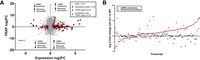
ADR-2-dependent gene expression changes are buffered at the translational level. (A) Quadrant scatter plot showing the fold change (log2) of germline expression (x-axis) and TRAP ribosome association (y-axis) in adr-2(-) germlines compared to wild type. Gray points denote genes with no significant change in either analysis. Red squares indicate a significant change in mRNA expression only (DESeq2 Padj ≤ 0.05, log2 fold change |≥0.5|) in adr-2(-) compared to wild type, and green triangles indicate a significant change in ribosome association only (DESeq2 Padj ≤ 0.05, log2 fold change |≥0.5|) in adr-2(-) compared to wild type. Blue diamond indicates a significant change in both expression and ribosome association (adr-2 only). DESeq2 analyses were performed with three biological replicates. Dotted lines represent log2 fold change significance cutoff. (B) Log2 fold change mRNA expression (red circles) and log2 fold change germline ribosome association (black squares) in adr-2(-) germlines compared to wild type for genes showing significant changes in mRNA expression (DESeq2 Padj ≤ 0.05, log2 fold change |≥0.5|).
To begin to distinguish between these possibilities, we sought to examine protein expression of genes that exhibit altered mRNA expression in germlines isolated from adr-2(-) animals compared to wild-type germlines. As antibodies specific to the C. elegans proteins are not readily available, we chose to make use of existing FLAG epitope-tagged strains of nas-37 and col-118, which exhibit 10- to 20-fold upregulated mRNA expression in germlines lacking adr-2 but similar levels of germline ribosome association in wild-type and adr-2(-) animals. Because both proteins were expressed at levels too low to be detected in isolated germlines (data not shown), quantitative immunoblotting was performed on whole young adult animals. Consistent with the translational buffering hypothesis, immunoblotting revealed similar levels of NAS-37 and COL-118 proteins in wild-type and adr-2(-) young adult animals (Supplemental Fig. S4). While additional proteins should be examined, particularly in isolated germlines, our data suggests that, similar to other germline studies of RNA binding protein mutants (Rochester et al. 2022), there are additional levels of secondary regulation, which can buffer changes in mRNA expression to maintain proper translational output in the absence of adr-2. This finding may also explain why loss of adr-2 alone, despite causing significant changes in gene expression at the mRNA level (Fig. 3C; Supplemental Table S3), does not cause any obvious defects in germline development or function. However, as several genes with associated germline and fertility phenotypes are misexpressed at the transcript level when adr-2 is absent, if factors necessary for the translational buffering mechanism are disrupted in this context, it seems likely that germline function and fertility would be affected. Interestingly, fertility defects have been observed when adr-2 is lost along with other factors (Reich et al. 2018; Fischer and Ruvkun 2020; Erdmann et al. 2024). While studies of germline gene expression regulators have demonstrated the presence of redundant and overlapping regulatory pathways that act as fail-safes to ensure proper germline function (Vanden Broek et al. 2022; Kelley et al. 2026), future studies specifically in adr-2(-) animals are needed to understand the mechanism of regulation at play here.
In sum, we demonstrate in this study that A-to-I RNA editing occurs at over 1000 sites across 71 germline transcripts. Interestingly, we see minimal changes in mRNA expression and translational status of edited transcripts in the absence of adr-2. While it is possible that editing has no effect on these transcripts under normal conditions, it is also possible that the effects of editing on these transcripts are not evident within the adult germline, where these experiments were performed. In addition to executing gametogenesis, a major function of the germline is to prepare for early embryonic development. As such, a cache of maternal transcripts is transcribed during gametogenesis and stored for use in early embryonic development before the onset of zygotic transcription (Rajyaguru and Parker 2009). Thus, editing of maternal transcripts in the germline may affect their fate in the embryo. While many of the germline edited transcripts identified in this study are known maternal transcripts (Supplemental Table S1; Quarato et al. 2021), further studies are needed to determine whether editing events are present on inherited early embryonic transcripts and whether editing impacts the stability or translation of these transcripts. It is also possible that the whole-germline nature of our studies has obscured effects on edited genes that may be restricted to specific cell types or stages in the germline. Single-cell studies and studies at different developmental stages may shed light on cell type– and stage-specific interactions and their potential effects on germline development and function.
Additionally, we demonstrate that adr-2 affects the expression of several ribosome components and rRNAs, leading to an overall reduction in both 18S and 26S rRNA concentrations. Studies of ribosome composition have revealed that ribosomes are highly heterogeneous, consisting of different sets of ribosomal proteins and rRNAs (Gay et al. 2022). Further studies have revealed that this heterogeneity can have functional consequences, with ribosomes of different composition preferentially translating different subsets of transcripts (Shi et al. 2017; Norris et al. 2021). Ribosome compositions have been shown to vary between tissues, developmental time points, and environmental conditions to facilitate unique translational regulation with functional consequences (Hopes et al. 2022; Li et al. 2022; Milenkovic and Novoa 2025). As such, the regulation of ribosome components by ADR-2 in the germline may contribute to germline-specific patterns of ribosome composition that ultimately regulate the germline translational landscape. While the TRAP assay performed in this study is useful for comparing translational efficiency of specific transcripts, its pulldown-based strategy makes it unsuitable for comparing bulk translational activity. More general activity assays, such as polysome profiling or puromycin incorporation assays, would be useful for determining whether regulation of ribosome components by adr-2 has a general effect on germline translational output. Future studies are needed to elucidate the molecular relationship between ADR-2, ribosome component expression, and germline translational activity, as well as the downstream consequences of ADR-2-dependent gene regulation on germline development and function.
MATERIALS AND METHODS
Worm strains and maintenance
All worm strains were maintained at 20°C on nematode growth media (NGM) seeded with Escherichia coli OP50. Worms were thawed regularly from frozen stocks to minimize effects of accumulated random mutations. The following previously generated strains were used in this study: Bristol Strain N2, SS104 (glp-4(bn2)) (Beanan and Strome 1992), BB19 (adr-1(tm668)) (Hundley et al. 2008), BB20 (adr-2(ok735)) (Hundley et al. 2008), BB21 (adr-1(tm668);adr-2(ok735)) (Hundley et al. 2008), SHG248 (control sensor) and SHG278 (risiRNA sensor) (Zhou et al. 2017), EV484 (efls155[mex-5p::rpl-4::FLAG::tbb-2 3′UTR + Cbr-unc-199(+)]II) (Nousch 2020), HAH36 (V5::adr-1;3xFLAG::adr-2) (Dhakal et al. 2024), HAH61 (3xFLAG::adr-2) (Dhakal et al. 2024), JDW436 (nas-37(wrd106[nas-37::mNG::3xFLAG])), and JDW827 (col-118(wrd343[col-118::mNG::3xFLAG])). Strains generated in this study include HAH87 (efls155[mex-5p::rpl-4::FLAG::tbb-2 3′UTR + Cbr-unc-199(+)]II), HAH88 (adr-2(ok735); efls155[mex-5p::rpl-4::FLAG::tbb-2 3'UTR + Cbr-unc-199(+)]II), HAH101 (control sensor), HAH102 (adr-2(-);control sensor), HAH103 (risiRNA sensor), HAH104 (adr-2(-); risiRNA sensor), HAH131 (nas-37(wrd106[nas-37::mNG::3xFLAG]), HAH132 (nas-37(wrd106[nas-37::mNG::3xFLAG]) adr-2(ok735)), HAH133 (col-118(wrd343[col-118::mNG::3xFLAG])), and HAH134 (col-118(wrd343[col-118::mNG::3xFLAG]) adr-2(ok735)).
Crossed strains were made by placing 10–15 males and one hermaphrodite on mating plates (NGM plates seeded with a small spot of E. coli OP50 in the center), and genotyping was performed for the F1 progeny and F2 progeny using primers mentioned in Supplemental Table S5. The specific crosses performed included creation of HAH87 and HAH88 by crossing EV484 hermaphrodites to BB20 males, creation of HAH101 and HAH102 by crossing SHG248 hermaphrodites to BB20 males, creation of HAH103 and HAH104 by crossing SHG278 hermaphrodites to BB20 males, creation of HAH131 and HAH132 by crossing JDW436 hermaphrodites to BB20 males, and HAH133 and HAH134 by crossing JDW827 hermaphrodites to BB20 males.
Bleaching
Synchronized animals were obtained by bleaching gravid adult animals with a solution of 5 M NaOH (33%) and sodium hypochlorite (Fisher Scientific) (66%). After the solution was added, animals were incubated on a shaker at 20°C for 7 min and then spun down to collect embryos. Embryos were washed with 1× M9 buffer (22 mM KH2PO4, 42.3 mM Na2HPO4, 85.6 mM NaCl, and 1 mM MgSO4) solution thrice and incubated overnight in 1× M9 buffer at 20°C. The next day, hatched L1 worms were spun down and washed again with 1× M9 buffer thrice.
Quantitative real-time PCR
RNA was extracted using standard TRIzol–chloroform extraction followed by treatment with TURBO DNase (Ambion) and cleanup with the RNeasy Extraction Kit (QIAGEN). Reverse transcription was performed on total RNA using random hexamer primers, oligo dT, and Superscript III (Thermo Fisher). Gene expression was determined using KAPA SYBR FAST Master Mix (Roche) and gene-specific primers (Supplemental Table S6) on a Thermo Fisher QuantStudio 3 instrument. The primers designed for qPCR spanned an exon–exon junction to prevent detection of genomic DNA in the samples. For each gene analyzed, a standard curve of eight to 10 samples of 10-fold serial dilutions of the amplified product was used to generate a standard curve of cycle threshold versus the relative concentration of amplified product. Standard curves were plotted on a logarithmic scale in relation to concentration and fit with a linear line. Fit (r2) values were around 0.99, and at least seven data points fell within the standard curve. Each cDNA measurement was performed in three technical replicates, and each experiment was performed in three biological replicates.
Germline immunostaining and imaging
Germlines were extruded from synchronized L4 or day-1 adult HH36 animals. Germlines were fixed with 1% paraformaldehyde and methanol and permeabilized with 0.2% Triton X-100 (Sigma-Aldrich). Germlines were stained with anti-V5 (Cell Signaling Technology) and anti-FLAG (Sigma-Aldrich) primary antibodies, followed by Alexa Fluor 594 (Thermo Fisher Scientific), Alexa Fluor 488 (Thermo Fisher Scientific), and DAPI (4′,6-diamidino-2-phenylindole) (Thermo Fisher Scientific). Germlines were mounted in Vectashield (Vector Labs) and imaged using a Leica SP8 Scanning Confocal Microscope (Indiana University Light Microscopy Imaging Center). Images were processed using ImageJ.
Germline sample collection for RNA-sequencing
Germline sample collection for RNA isolation was performed as previously described (Campbell and Updike 2015), with slight modifications. Synchronized young adult (60 h post egg-lay) animals were used for germline dissection. Animals were picked into egg buffer (118 mM NaCl, 48 mM KCl, 2 mM CaCl2, 2 mM MgCl2, 25 mM HEPES, pH 7.3 adjusted osmolarity to 340 mOsm with sucrose) containing levamisole (2.5 mM). Animals were cut at the pharynx to allow for extrusion of the gonad. Germlines were collected via mouth aspiration using a pulled glass capillary coated with Sigmacote (Sigma-Aldrich). For germline RNA samples, germlines were dispensed into 400 µL TRIzol on ice and then frozen in liquid nitrogen. RNA was extracted using standard TRIzol–chloroform extraction followed by treatment with TURBO DNase (Ambion) and cleanup with the RNeasy Extraction Kit (QIAGEN).
RNA-sequencing library preparation
For both the germline RNA-seq experiments and the TRAP-seq experiment: DNase-treated total RNA was subjected to two rounds of poly(A) selection using magnetic oligo(dT) beads (Invitrogen). Libraries were created from entire poly(A)-selected RNA samples using the KAPA Stranded RNA-seq Library Prep kit (Roche); cleanup steps were performed using AMPure XP beads (Beckman Coulter). Library fragment size distribution (200–900 bp) was determined via TapeStation electrophoresis (Agilent). Libraries for each of three biological replicates per condition were pooled by concentration and sequenced on a NextSeq 2000 (75 cycles) flow cell at the Indiana University Center for Genomics and Bioinformatics. The quality of the sequencing reads obtained was checked using FASTQC (version 0.12.1), the results of which are summarized in Supplemental Table S6.
Bioinformatic analysis of germline RNA-sequencing data
Reads were aligned to a reference genome (C. elegans PRJNA13758 WS275) using STAR (version 2.7.10b). Percent uniquely mapped reads for each sample are listed in Supplemental Table S5. A-to-I editing site identification was performed using SAILOR (Deffit et al. 2017; Kofman et al. 2023). Each biological replicate was analyzed separately. Editing sites called at ≥75% confidence in all three replicates of wild-type germlines were selected as high-confidence editing sites. Editing sites also called in adr-2(-) germlines (regardless of confidence) were removed as false positives (Supplemental Fig. S5). Variant analysis was performed using BCFtools mpileup (Danecek et al. 2021). Differential gene expression analyses were performed in R with DESeq2 (Love et al. 2014). Gene ontology enrichment analyses were performed with WormCat (Holdorf et al. 2020). Full code for these analyses is available at https://github.com/emierdma/GSF3037.
Two logo analysis
Germline- and neural-specific edit sites and their genomic coordinates were extracted from Supplemental Table S1 and converted to BED files with strand information pulled from the reference genome (C. elegans PRJNA13758 WS275). Nucleotides flanking the edit sites (±2 nt) were extracted with BEDTools (Quinlan and Hall 2010), and the resulting 5mers were converted to FASTA. Control samples were comprised of a random adenosine and its ±2 flanking nucleotides that were ∼2000 nt away from edit sites. Control samples matched experimental samples in number of sites. Two Sample Logo (http://twosamplelogo.org) was used to identify under- and overrepresented flanking nucleotides.
Germline sample collection and rRNA measurement
Germlines were dissected for RNA isolation as described above. An equal amount of a spike-in RNA of known concentration (C. elegans lam-2 synthesized via in vitro transcription, adapted from Rajendren et al. 2018) was added to each germline sample, and then RNA was extracted using standard TRIzol–chloroform extraction followed by treatment with TURBO DNase (Ambion) and cleanup with the RNeasy Extraction Kit (QIAGEN). Total RNA was run on the Agilent TapeStation using High Sensitivity RNA ScreenTape (Agilent). Peaks for the spike-in RNA (160 nt), 18S rRNA (∼1000 nt), and 26S rRNA (∼2000 nt) were automatically assigned by the TapeStation software, with adjustments as needed. Concentrations for spike-in, 18S rRNA, and 26S rRNA were calculated by the TapeStation software.
Translating ribosome affinity purification (TRAP)
Animals were synchronized via bleaching and allowed to grow to young adulthood. Young adult animals were washed from plates with 1× M9 buffer and incubated for 20 min without food to allow for digestion of OP50. Animals were then washed once in extract buffer (50 mM HEPES, 70 mM K-acetate, 5 mM Mg-acetate, 10% glycerol, 1% NP-40, pH 7.4) with added EDTA-free protease inhibitor (Roche) and cycloheximide (100 μg/mL) and resuspended in an equal volume of concentrated extract buffer with protease inhibitor and cycloheximide before being dripped into liquid nitrogen to make pellets. Pellets were ground to a fine powder and allowed to thaw and then centrifuged to generate a clear lysate. Input lysate was taken and either boiled with 6× SDS buffer for 5 min for western blotting or frozen in 400 µL TRIzol for RNA isolation. For each immunoprecipitation (IP), 200 µg of lysate was added to 25 µL of prewashed FLAG magnetic resin along with 200 µL of concentrated extract buffer with cycloheximide and brought up to 1 mL volume with extract buffer with no added detergents. Lysates were incubated with resin for 2 h at 4°C and then washed thrice with KCl wash buffer (10 mM HEPES pH 7.4, 150 mM KCl, 5 mM MgCl2, and 1% NP-40) with added cycloheximide (100 μg/mL) and protease inhibitor (Roche) and twice with 1× TBS. For IPs taken for western blotting, beads were resuspended in 40 µL 2× SDS buffer and boiled for 5 min. For IPs taken for RNA isolation, beads were resuspended in 1× TBS and incubated with 0.5 µL Proteinase K for 15 min at 42°C with shaking at 1200 rpm. Supernatant was removed from beads and added to 400 µL TRIzol and frozen.
Western blotting
For TRAP experiments, input and IP samples were run on an SDS-PAGE gel and then transferred to a nitrocellulose membrane. For the whole worm analyses of COL-118 and NAS-37 expression, lysates from young adult animals were run on an SDS-PAGE gel and then transferred to a nitrocellulose membrane. Membranes were blocked in milk for 1 h and then incubated in 1× TBS containing primary antibody (1:2000 αFLAG (Sigma-Aldrich F1804), (1:1000 αβ-actin [Cell Signaling Technologies D6A8]) overnight at 4°C. Membranes were washed thrice with 1× TBS and then incubated in 1× TBS containing secondary antibody (FLAG: 1:40,000 αMouse HRP [TRAP] or 1:20,000 αMouse HRP (NAS-37, COL-118; actin: 1:40,000 αRabbit HRP)) for 1 h. Membranes were washed thrice more in 1× TBS before developing with ECL (either 50% pico/50% femto [TRAP] or 100% pico [NAS-37/COL-118 experiments], Fisher). Blots were imaged on a Bio-Rad ChemiDoc instrument. Band intensities were quantified using ImageJ.
Bioinformatic analysis of TRAP-sequencing data
Reads were aligned to a reference genome (C. elegans PRJNA13758 WS275) using STAR (version 2.7.11a). Percent uniquely mapped reads for each sample are listed in Supplemental Table S6. Differential gene expression analyses were performed in R with DESeq2 (Love et al. 2014). Full code for these analyses can be found at https://github.com/emierdma/GSF3037.
DATA DEPOSITION
Strains and plasmids are available upon request. Raw and processed high-throughput sequencing data generated in this study have been submitted to the NCBI Gene Expression Omnibus (GEO; https://www.ncbi.nlm.nih.gov/geo/) under accession number GSE299666 and to the EMBL-EBI European Nucleotide Archive (ENA; https://www.ebi.ac.uk/ena/browser/home) under accession numbers PRJEB100883 and PRJEB100892.
SUPPLEMENTAL MATERIAL
Supplemental material is available for this article.
ACKNOWLEDGMENTS
We thank Dustin Updike for his guidance with C. elegans germline isolation for RNA sequencing, the Indiana University Center for Genomics and Bioinformatics for running the high-throughput sequencing experiments, and the Indiana University Light Microscopy Imaging Center for the use of the Leia SP8 Confocal. This work was supported in part by the National Institutes of Health/National Institute of General Medical Sciences (R35GM156459) awarded to H.A.H., the National Institutes of Health/National Institute of General Medical Sciences (T32GM131994), and the National Institutes of Health/National Institute of Child Health and Human Development (F31HD110244) awarded to E.A.E. Some strains were provided by the Caenorhabditis Genetics Center (CGC), which is funded by the NIH Office of Research Infrastructure Programs (P40OD010440).
Footnotes
-
Article is online at http://www.rnajournal.org/cgi/doi/10.1261/rna.080820.125.
-
Freely available online through the RNA Open Access option.
- Received October 16, 2025.
- Accepted February 25, 2026.
This article, published in RNA, is available under a Creative Commons License (Attribution 4.0 International), as described at http://creativecommons.org/licenses/by/4.0/.




