


Thioredoxin regulates the redox state and the activity of the human tRNA ligase complex
- 1Max Perutz Laboratories, Medical University of Vienna, Vienna Biocenter (VBC), 1030 Vienna, Austria
- 2Vienna Biocenter PhD Program, a Doctoral School of the University of Vienna and Medical University of Vienna, 1030 Vienna, Austria
- Corresponding author: javier.martinez{at}meduniwien.ac.at
Abstract
The mammalian tRNA ligase complex (tRNA-LC) catalyzes the splicing of intron-containing pre-tRNAs in the nucleus and the splicing of XBP1 mRNA during the unfolded protein response (UPR) in the cytoplasm. We recently reported that the tRNA-LC coevolved with PYROXD1, an essential oxidoreductase that protects the catalytic cysteine of RTCB, the catalytic subunit of the tRNA-LC, against aerobic oxidation. In this study, we show that the oxidoreductase Thioredoxin (TRX) preserves the enzymatic activity of RTCB under otherwise inhibiting concentrations of oxidants. TRX physically interacts with oxidized RTCB, and reduces and reactivates RTCB through the action of its redox-active cysteine pair. We further show that TRX interacts with RTCB at late stages of UPR. Since the interaction requires oxidative conditions, our findings suggest that prolonged UPR generates reactive oxygen species. Thus, our results support a functional role for TRX in securing and repairing the active site of the tRNA-LC, thereby allowing pre-tRNA splicing and UPR to occur when cells encounter mild, but still inhibitory levels of reactive oxygen species.
INTRODUCTION
RNA molecules are mostly synthesized as precursors and only become functional upon a series of modifications and processing events such as the removal of intronic sequences during a process called RNA splicing. The so-called “canonical splicing” entails the removal of introns from precursor mRNAs (pre-mRNAs) (Berget et al. 1977; Chow et al. 1977). In contrast, the term “noncanonical splicing” has been coined to describe the removal of single introns from pre-tRNAs (Hopper and Banks 1978; Knapp et al. 1978, 1979; O'Farrell et al. 1978; De Robertis and Olson 1979; Peebles et al. 1979, 1983; Schmidt and Matera 2020; for review, see Phizicky and Hopper 2023).
Pre-tRNA splicing occurs in two steps: cleavage of the pre-tRNA by a tRNA splicing endonuclease (Ho et al. 1990; Rauhut et al. 1990; Trotta et al. 1997; Paushkin et al. 2004; Hayne et al. 2023; Sekulovski et al. 2023; Zhang et al. 2023) and ligation of the resulting tRNA exon halves by an RNA ligase to generate a mature tRNA (Filipowicz and Shatkin 1983; Greer et al. 1983; Laski et al. 1983; Phizicky et al. 1986). In mammals, the ligation of tRNA exon halves is executed by the tRNA ligase complex (tRNA-LC), composed of the catalytic subunit RTCB (RNA-splicing ligase RtcB homolog) and four additional subunits: DDX1, CGI-99 (RTRAF/hCLE), FAM98B, and Ashwin (ASW/C2orf49) (Popow et al. 2011, 2012). The tRNA-LC becomes a multiple turnover enzyme through guanylylation by Archease (Popow et al. 2014; Desai et al. 2015). Splicing of intron-containing pre-tRNAs is essential because specific tRNA isodecoder families are exclusively encoded as intron-containing pre-tRNAs and therefore need to be spliced in order to be functional in protein translation (Chan and Lowe 2016; Gogakos et al. 2017).
Another example of noncanonical splicing is the cytoplasmic removal of a retained intron from an mRNA during the unfolded protein response (UPR), a signaling cascade that is triggered when unfolded proteins accumulate in the ER (Kozutsumi et al. 1988; Walter and Ron 2011). This pathway aims to reestablish ER homeostasis to ensure survival of the cell, but can also induce apoptosis if activated over a prolonged period of time. The most conserved branch of the UPR comprises IRE1, an RNA endonuclease and kinase, that cleaves the retained intron in the XBP1u mRNA (u for unspliced) upon activation (Sidrauski and Walter 1997; Yoshida et al. 2001). In metazoans, the cleaved XBP1 mRNA exons are joined by the tRNA-LC to generate XBP1s mRNA (s for spliced) (Jurkin et al. 2014; Kosmaczewski et al. 2014; Lu et al. 2014). Translation of this mRNA results in the transcription factor XBP1s, which up-regulates proteins that assist in resolving the UPR (Lee et al. 2003; Acosta-Alvear et al. 2007). Besides these well-studied RNA ligation reactions, the tRNA-LC is required for the splicing of transcripts related to DNA methylation and DNA damage repair in Mus musculus (Zhang et al. 2022).
We have recently reported that the mammalian tRNA-LC is reversibly inhibited by oxidative stress (Asanovic et al. 2021). Structural studies showed that the cysteine in the active site of an archaeal RTCB is prone to oxidation (Banerjee et al. 2021), suggesting that the inactivation of the mammalian tRNA-LC likely also occurs through oxidation of the catalytically active cysteine. Furthermore, we have shown that the oxidoreductase PYROXD1 coevolved with the tRNA-LC to protect its catalytic center against aerobic oxidation, but not to rescue the tRNA-LC once oxidized (Asanovic et al. 2021). How the oxidized and inactivated tRNA-LC is reactivated remains unknown, as much as the physiological relevance of its regulation by a redox mechanism. Reduction of oxidized proteins during recovery from oxidative stress in cells is usually achieved by oxidoreductases such as Thioredoxin (TRX) (Luthman and Holmgren 1982; Lee et al. 2014). TRX and its homologs, such as the mitochondrial TRX2, reduce their target proteins using two catalytic cysteines—C32 and C35 in humans—within the CxxC motif (Lee et al. 2013). A proteome-wide study aiming to find targets of mammalian TRX and TRX-like proteins identified RTCB among other targets (Nakao et al. 2015). However, it remains unclear if this interaction has any physiological relevance.
In this study, we show that the absence of TRX enhances the sensitivity of the tRNA-LC toward oxidative stress, with an impact on pre-tRNA and XBP1 mRNA splicing. We demonstrate that TRX reactivates the tRNA-LC after alleviation of the oxidative stress as well as upon extended UPR through direct interaction with RTCB.
RESULTS AND DISCUSSION
TRX increases the tolerance of the tRNA ligase complex to oxidative stress
To test whether TRX preserves the enzymatic activity of the human tRNA-LC during oxidative stress, we generated a HeLa cell line expressing a Doxycycline (Dox) inducible short hairpin RNA (shRNA) targeting the TXN mRNA encoding TRX (shTRX cells) (Zuber et al. 2011). Both TRX protein and TXN mRNA levels decreased after 3 d of shRNA expression, while RTCB protein levels remained unchanged (Supplemental Fig. 1A,B). We monitored tRNA-LC activity through the interstrand ligation of a double-stranded, radiolabeled RNA substrate bearing a 3′phosphate (3′P) on one strand and a 5′hydroxyl group (5′OH) on the complementary strand (Fig. 1A; Popow et al. 2011). Under standard conditions, extracts from shTRX cells and control shRNA cells (shCtrl cells) displayed similar ligation activity (Fig. 1A,B). However, upon treatment with increasing concentrations of the oxidant menadione, the tRNA-LC became inactive at lower concentrations of menadione in shTRX cells (Fig. 1C,D). We calculated IC50 values of 27 µM in shCtrl cells and 18 µM in shTRX cells. Treating cells with increasing concentrations of H2O2 caused a similar effect (Supplemental Fig. 1C,D), with IC50 values of 76 µM for shCtrl cells and 37 µM for shTRX cells. RTCB levels remained unchanged in both treatments (Fig. 1E; Supplemental Fig. 1E). Of note, we observed an increase in the activity of the tRNA-LC irrespective of the presence of TRX at 5 and 10 µM menadione, but not in the presence of H2O2. This effect may relate to other conserved cysteine residues in the subunits of the tRNA-LC which are potentially modified during oxidative stress. Yet, the absence of TRX has a clear impact on the sensitivity of the tRNA-LC in the presence of menadione and H2O2.
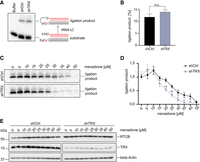
Depletion of TRX by shRNAs sensitizes the tRNA-LC to oxidative stress. (A) HeLa cells expressing a short RNA hairpin (shRNA) targeting TXN mRNA to deplete TRX protein (shTRX), or a control shRNA targeting Renilla luciferase (shCtrl) upon addition of 2 µg/mL Dox, were lysed after 3 d of shRNA expression and RNA ligation activity was assayed by incubating cell lysates with a double-stranded, radiolabeled RNA substrate containing a 3′ phosphate on one strand and a 5′OH on the complementary strand. Products of the reaction were resolved by denaturing urea polyacrylamide gel electrophoresis and visualized by phosphorimaging. (B) Quantification of band intensities from A. The substrate conversion rate was calculated as the quotient of the substrate and the sum of substrate and product band intensities. (n = 7, mean values ± SEM are shown, significances were analyzed using unpaired Student's t-test assuming unequal variances, with [*] P < 0.05; [**] P < 0.01; [***] P < 0.005). (C) ShCtrl and shTRX cells were treated with increasing concentrations of menadione for 1 h before harvest and lysis. Cell lysates were assayed as in A. (D) Quantification of band intensities from C. The substrate conversion rate was calculated as in B, with n = 6. (E) Levels of RTCB and TRX were assessed in shTRX and shCtrl cell lysates after treatment with increasing concentrations of menadione for 1 h by western blot using anti-RTCB and anti-TRX antibodies. Levels of β-Actin were used as a loading control.
Catalytically active TRX is required to sustain tRNA-LC activity during oxidative stress and to reactivate the tRNA-LC once the stress is mitigated
We next evaluated whether the catalytic cysteine residues of TRX are required to support tRNA-LC activity during oxidative stress. We therefore overexpressed shRNA-resistant, FLAG-tagged variants of human TRX—FLAG-TRX CxxC (WT) and FLAG-TRX SxxS (mutant)—in shTRX cells (Supplemental Fig. 2A,B). To note, mutating the active site of TRX affected the recognition of FLAG-TRX by the anti-TRX antibody but not by the anti-FLAG antibody, indicating a similar level of overexpression of the constructs. Overexpression of FLAG-TRX CxxC prevented the inactivation of the tRNA-LC by menadione (Fig. 2A,B), while overexpression of FLAG-TRX SxxS did not (Fig. 2A,B). This shows that the catalytic cysteines of TRX are required to sustain the enzymatic activity of the tRNA-LC during oxidative stress.
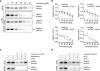
Catalytically active TRX is required to maintain tRNA-LC activity during oxidative stress and for its reactivation after oxidative stress levels decrease. (A) ShRNA-resistant wild-type FLAG-TRX CxxC (labeled as CxxC) or active site double mutant FLAG-TRX SxxS (labeled as SxxS) were constitutively overexpressed in shTRX cells. Silencing of endogenous TRX was induced by the addition of 2 µg/mL Dox for 3 d. Cells were treated with menadione for 1 h before harvest and lysis, and RNA ligation activity was assayed by incubating cell lysates with a double-stranded, radiolabeled RNA substrate containing a 3′ phosphate on one strand and a 5′OH on the complementary strand. Products of the reaction were resolved by denaturing urea polyacrylamide gel electrophoresis and visualized by phosphorimaging. (B) Quantification of band intensities from A. The substrate conversion rate was calculated as the quotient of the substrate and the sum of substrate and product band intensities and normalized to untreated shCtrl sample (n = 3, mean values ± SEM are shown. Significances were analyzed using unpaired Student's t-test assuming unequal variances, with [*] P < 0.05; [**] P < 0.01; [***] P < 0.005). Top left graph: comparison of ligation product in shCtrl versus shTRX FLAG-TRX CxxC cells. Top right graph: comparison of ligation product in shCtrl versus shTRX FLAG-TRX SxxS cells. Bottom left graph: comparison of ligation product in shTRX versus shTRX FLAG-TRX CxxC cells. Bottom right graph: comparison of ligation product in shTRX versus shTRX FLAG-TRX SxxS cells. (C) HeLa cells were transfected with siRNA pools targeting TXN mRNA (siTRX) or a control siRNA (siCtrl) for 3 d. Cells were treated with 30 µM menadione for 30 min, washed, and allowed to recover for 0, 1, or 3 h in medium containing 10 µg/mL CHX. As a control, cells were either left untreated or treated with CHX alone for 5 h. Cells were lysed and assayed for RNA ligation as in A. (D) HeLa cells were transfected with siRNA pools targeting TXN mRNA (siTRX) or a control siRNA (siCtrl) for 3 d. Cells were treated with 125 µM H2O2 for 30 min, washed, and allowed to recover for 0, 1, or 3 h in medium containing 10 µg/mL CHX. As a control, cells were either left untreated or treated with CHX alone for 5 h. Cells were lysed and assayed for RNA ligation as in A.
We next investigated the potential role of TRX in the previously described reversibility of the tRNA-LC inhibition upon recovery from oxidative stress (Asanovic et al. 2021), and in the reactivation of the inhibited tRNA-LC. These experiments were performed in HeLa cells transiently transfected with siRNAs, instead of the Dox-inducible shRNA system, to avoid the antioxidant effect triggered by Dox (Clemens et al. 2018), which can affect the reactivation phase. We first performed titrations to determine the minimal inhibitory concentrations of menadione and H2O2 in siCtrl and siTRX cells. We observed an almost complete inhibition of tRNA-LC activity at 30 µM menadione in siCtrl cells, while tRNA-LC activity was abrogated at 20 µM menadione in siTRX cells (Supplemental Fig. 2C). Inhibition with H2O2 was achieved at 250 µM in siCtrl cells, while 100 µM substantially reduced RNA ligation activity in siTRX cells (Supplemental Fig. 2D). To monitor the reactivation of the tRNA-LC after oxidative stress, we treated cells with 30 µM menadione, while further titrations with H2O2 led us to an optimal concentration of 125 µM H2O2. The treatment was done for 30 min and cells were allowed to recover over a time course of 3 h in medium supplemented with cycloheximide (CHX) to inhibit de novo protein synthesis. In siCtrl cells treated with menadione, the tRNA-LC fully recovered after 3 h (Fig. 2C). In contrast, we did not detect tRNA-LC activity in siTRX cells (Fig. 2C). Similar to menadione treatment, we were not able to detect tRNA-LC activity in siTRX cells treated with 125 µM H2O2 (Fig. 2D). Importantly, levels of RTCB did not change during the recovery period after menadione or H2O2 treatment (Supplemental Fig. 2E,F). These results indicate that catalytically active TRX is required to reactivate the tRNA-LC once oxidative stress levels have decreased.
RTCB, the catalytic subunit of the tRNA-LC, is a substrate of TRX
We performed kinetic trapping pulldowns to test whether the reductive function of TRX relies on a physical interaction with the tRNA-LC (Nakao et al. 2015). We overexpressed FLAG-TRX CxxS in cells depleted of endogenous TRX to stabilize a potential interaction between the tRNA-LC and FLAG-TRX CxxS. Through mutation of the resolving C35, targets of TRX that have an oxidized cysteine residue (e.g., −SOH or a disulfide bond between two cysteines) are “trapped” due to the formation of an intermediate disulfide bond that cannot be reduced (Fig. 3A). We immunoprecipitated FLAG-TRX CxxS from untreated cells and from cells treated with different concentrations of H2O2 for 3 min, and carried out elution with FLAG-peptide in nonreducing conditions. We found RTCB enriched in the eluate in a H2O2 concentration-dependent manner, indicating that RTCB is a target of TRX (Fig. 3B). Peroxiredoxin 1 (PRDX1), a well-known target of TRX (Chae et al. 1999), was also detected in the eluate. A similar enrichment for RTCB and PRDX1 was observed in cells treated with increasing concentrations of menadione (Fig. 3C). We could not detect other subunits of the tRNA-LC in eluates of H2O2- or menadione-treated cells (Fig. 3D). As expected, trapping was neither detected for FLAG-TRX CxxC nor for FLAG-TRX SxxS mutants upon treatment with H2O2 (Fig. 3E).
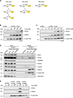
RTCB is a substrate of TRX. (A) Scheme of the reduction mechanism of TRX CxxC toward its target protein containing an oxidized cysteine residue (left panel). Trapping of target proteins using TRX CxxS (middle panel). The double mutant SxxS cannot reduce the oxidized cysteine on the target protein due to mutation of the catalytic cysteines (right panel). (B) shTRX cells constitutively overexpressing FLAG-TRX CxxS were treated for 3 min with increasing concentrations of H2O2 before lysis. ShTRX cells were left untreated as a negative control. Immunoprecipitation and elution were performed in nonreducing conditions. Eluates were analyzed by western blot using anti-RTCB, anti-PRDX1, and anti-TRX antibodies. (C) Trapping was performed as described in B after treatment with increasing concentrations of menadione for 15 min. ShTRX cells were left untreated as a negative control. (D)Trapping was performed as described in B in shTRX cells expressing FLAG-TRX CxxS after treatment with 100 µM H2O2 for 3 min or 40 µM menadione for 15 min. Untreated shTRX and shTRX overexpressing FLAG-TRX CxxS were used as controls. Inputs and eluates were analyzed by western blot using anti-DDX1, anti-RTCB, anti-FAM98B, anti-CGI-99, anti-PRDX1, and anti-TRX antibodies. (E) Trapping was performed as described in B in shTRX cells expressing FLAG-TRX CxxC, FLAG-TRX CxxS, and FLAG-TRX SxxS after treatment of cells with 0 or 100 µM H2O2 for 3 min.
Thus, our data demonstrate that TRX directly interacts with RTCB in an oxidant-dependent manner. This suggests that the increased redox sensitivity of the tRNA-LC in the absence of TRX, as well as the reversible character of the inactivation, is a consequence of the interaction between TRX and RTCB. Furthermore, these results strongly imply that the reactivation of RTCB is through the reductive function of TRX, presumably by reduction of the active site cysteine of RTCB. This is in line with the structural study from Pyrococcus horikoshii, where oxidation of the active site cysteine was observed in aerobically purified RTCB (Banerjee et al. 2021). Oxidation of the highly conserved cysteine (C122 in humans) causes loss of metal ion binding and therefore loss of catalytic activity. We hypothesize that TRX can reduce C122 and thereby allows reloading of the active site with the metal ions to regain its function.
Depletion of TRX during oxidative stress impairs pre-tRNA splicing and XBP1 mRNA splicing during UPR
We tested the TRX-RTCB interplay during pre-tRNA splicing and XBP1 mRNA splicing in conditions of oxidative stress. For this purpose, we assayed pre-tRNA splicing in vitro using a radiolabeled, intron-containing pre-tRNA from Saccharomyces cerevisiae and lysates from shCtrl and shTRX cells treated with menadione. Cleavage of the substrate pre-tRNA with the consequent generation of tRNA exon halves was not impaired by oxidative stress (Fig. 4A, bottom panels; Asanovic et al. 2021). However, ligation of exon halves to generate a mature tRNA was inhibited at 25 µM menadione in shTRX cells, compared to 50 µM menadione in shCtrl cells (Fig. 4A, top panels).
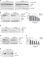
Depletion of TRX impairs the physiological functions of the tRNA-LC. (A) ShCtrl and shTRX cells were treated with indicated menadione concentrations for 1 h before harvest and lysis. An internally radiolabeled S. cerevisiae pre-tRNA substrate was added to cell lysates to monitor cleavage by the TSEN complex and ligation to mature tRNAs by the tRNA-LC. Pre-tRNA processing was monitored by denaturing urea gel electrophoresis and visualized by phosphorimaging. (B) ShCtrl and shTRX were treated with menadione for 1 h. RNA was isolated and subjected to northern blot analysis using a probe complementary to the 5′exon of Tyr-tRNA. A probe targeting U6 snRNA was used as a loading control. (C) ShCtrl and shTRX cells were treated with indicated menadione concentrations and 1.5 µg/mL Tm for 4 h before RNA isolation and cDNA synthesis. RT-qPCR was performed to measure XBP1s and XBP1u mRNA levels relative to ACTB (shown as the ratio XBP1s/XBP1u). (n = 9, mean values ± SEM are shown. Significances were analyzed using paired Student's t-test assuming unequal variances, with [*] P < 0.05; [**] P < 0.01; [***] P < 0.005). (D) ShCtrl and shTRX cells were treated with indicated menadione concentrations and 1.5 µg/mL Tm for 4 h before RNA isolation and cDNA synthesis. Semiquantitative RT-PCR was performed to analyze levels of XBP1 mRNA and ACTB mRNA. PCR products were resolved by agarose gel electrophoresis. (E) ShCtrl and shTRX cells were treated with indicated menadione concentrations and 1.5 µg/mL Tm for 4 h before harvest. Cells were lysed and protein levels of XBP1s and TRX were analyzed by western blot using respective antibodies. β-Actin levels were assessed as a loading control. (F) HEK293 FITR cells expressing GFP-XBP1intron-mCherry were transfected with siRNA pools targeting TXN (siTRX) or control siRNA (siCtrl) for 3 d. Expression of the reporter was induced for 5 h with 2 µg/mL Dox and treatment with menadione, and 1.5 µg/mL Tm was performed for 4 h. Splicing of the intron (generation of GFP-mCherry fusion protein) was assessed by FACS analysis. The graph only shows GFP-mCherry positive cells (n = 4, mean values ± SEM are shown. Significances were analyzed using unpaired Student's t-test assuming unequal variances, with [*] P < 0.05; [**] P < 0.01; [***] P < 0.005). (G) ShTRX FLAG-TRX CxxS cells were treated for 4 or 8 h with 1.5 µg/mL Tm, or for 4 or 8 h with 300 nM Tg before lysis. As a positive control for trapping, shTRX FLAG-TRX CxxS cells were treated for 3 min with 100 µM H2O2. ShTRX cells were left untreated as a negative control. Immunoprecipitation and elution were performed in nonreducing conditions. Eluates were analyzed by western blot using anti-RTCB or anti-TRX antibodies.
To further validate the protective role of TRX toward the tRNA-LC under oxidative stress conditions, we performed a northern blot analysis to detect a fragment of Tyr-tRNA, composed of the 5′ leader sequence followed by the 5′ exon, that accumulates upon inhibition of the tRNA-LC (Hanada et al. 2013). The Tyr-tRNA fragment was detected at lower concentrations of menadione in shTRX cells compared to shCtrl cells (Fig. 4B). Of note, levels of total pre-tRNAs after treatment with 40 and 50 µM menadione were reduced in both cell lines, possibly due to Pol III inhibition (Gouge et al. 2015).
We next monitored XBP1 mRNA splicing upon induction of UPR with Tunicamycin (Tm) and simultaneous oxidative stress conditions. The splicing efficiency, determined by the ratio of XBP1s to XBP1u mRNA, decreased in shCtrl and shTRX cells after menadione treatment when measured by RT-qPCR (Fig. 4C). However, in shTRX cells, 30 µM menadione was sufficient for a significant reduction in comparison to shCtrl cells, where treatment with 40 µM menadione was required. A similar effect was observed when measuring levels of XBP1u and XBP1s mRNA by semiquantitative RT-PCR (Fig. 4D). We also analyzed levels of XBP1s protein by western blot and observed a reduction at 40 µM menadione in shTRX cells, while 50 µM menadione was required in shCtrl cells to achieve a similar effect (Fig. 4E). The reduction in XBP1s mRNA and XBP1 protein levels in the absence of TRX and simultaneous oxidative stress also affected XBP1s target genes, as shown by the reduction of DNAJB9 mRNA in shTRX cells treated with 30 µM menadione in comparison with shCtrl cells (Supplemental Fig. 3A).
We furthermore developed a FACS-based XBP1 mRNA splicing reporter in a HEK293 FITR cell line. The reporter encodes a Dox-inducible, GFP-XBP1intron-mCherry transcript that generates a GFP-mCherry fusion protein upon splicing, similar to previously described XBP1 splicing reporters (Lu et al. 2014; Roy et al. 2017). First, we validated the functionality of the reporter by codepleting RTCB and Archease, which was shown to be required for a decrease in XBP1s generation (Jurkin et al. 2014). We observed a drastic reduction in the generation of GFP-mCherry when UPR was induced with Thapsigargin (Tg) in cells depleted of RTCB and Archease, demonstrating that the reporter indeed reflects endogenous XBP1 mRNA splicing (Supplemental Fig. 3B). When TRX was depleted using siRNAs (siTRX cells) and UPR was induced simultaneously with oxidative stress, the levels of GFP-mCherry protein diminished at lower menadione concentrations in comparison to siCtrl cells (Fig. 4F). The difference in the concentrations of menadione required to observe a decrease in the expression of the GFP-mCherry reporter and the endogenous XBP1 protein in shCtrl/shTRX cells is due to the use of different cell lines: HEK293 FITR for the reporter and HeLa cells for the shRNA-mediated silencing. Taken together, these results demonstrate that TRX is required to maintain the physiological activity of the tRNA-LC when cells encounter oxidative stress.
We finally took advantage of the sensitive trapping approach to test whether the physical interaction between TRX and the tRNA-LC also occurs as a consequence of ROS generated endogenously during UPR. We therefore treated cells for 4 or 8 h with Tm or Tg to induce UPR. As a positive control for the trapping, we treated cells with 100 µM H2O2 (Fig. 3). We detected a robust interaction between FLAG-TRX CxxS and RTCB when treating cells with Tg for 8 h (Fig. 4G). This result suggests that ROS are generated during prolonged UPR and that RTCB is supported by the reductive activity of TRX.
The tRNA-LC is able to catalyze the splicing of XBP1 mRNA at low concentrations of exogenous ROS in the presence of TRX (Fig. 4C–F); however, TRX cannot cope with higher levels of oxidation, leading to a decrease in XBP1 mRNA splicing and, consequently, in the expression of XBP1s target genes (Supplemental Fig. 3A). Thus, potentially higher levels of ROS generated at late stages of UPR could irreversibly inhibit the tRNA-LC, down-regulate XBP1 mRNA splicing and signal to induce apoptosis.
This study demonstrates that the redox sensitivity of the tRNA-LC is a tightly regulated process that involves several factors and impacts pre-tRNA splicing, UPR and ultimately, protein biosynthesis. PYROXD1 has been shown to protect RTCB from oxidation (Asanovic et al. 2021) and, recently, to dissociate from RTCB before substrate binding (Loeff et al. 2023). Such dissociation offers an opportunity for ROS to oxidize RTCB. We envision that TRX acts at this step, bringing RTCB back to a functional state.
MATERIALS AND METHODS
Cell culture
HeLa Kyoto, HeLa RIEP (Jurkin et al. 2014) and HEK293 Flip-In T-REx (FITR, Invitrogen) cells were cultured in Dulbecco's modified Eagle's medium (DMEM high glucose, Gibco, #41966-052) supplemented with 10% fetal bovine serum (FBS, Gibco, #A5256701), 100 µg/mL penicillin/100 µg/mL streptomycin (Sigma Aldrich, #P0781-100ml), and 20 mM HEPES pH 7.0 (Gibco, #15630-122), at 37°C, 5% CO2. Cells were regularly tested for mycoplasma contamination.
Generation of Dox-inducible shRNA cells lines and induction of shRNA expression
For generation of Doxycycline-inducible shRNA cell lines, HeLa RIEP cells were transduced with ecotropically packaged shRNA expression vectors (pSIN-TRE3G-GFP-miRE-PGK-Neo, see “Cloning of shRNAs”) or rescue vectors (pBMN-FLAG-IRES-GFP, see “Cloning of shRNA-resistant TRX-rescue constructs”), as described before (Fellmann et al. 2013). For retroviral packaging, Platinum E cells (PlatE, Cell Biolabs), were seeded into 10 cm2 plates and transfected at 90% confluency with 20 µg plasmid DNA and 10 µg helper plasmid (pCMV-Gag-Pol, Cell Biolabs), using CalPhos Mammalian Transfection Kit (Clontech/Takara, #631312), according to the manufacturer's instructions. The transfection medium was replaced to complete DMEM after 16 h. Virus-containing supernatants were harvested at 36, 48, and 60 h post-transfection and added dropwise onto HeLa RIEP cells for transduction. Cells expressing the shRNA constructs were selected using 1 mg/mL Neomycin (Calbiochem, #480100). To induce expression of shRNAs, cells were treated for 3 d with 2 µg/mL Dox.
XBP1 splicing reporter
The XBP1 splicing reporter was designed similar to previously described studies (Lu et al. 2014). A 120-mer oligonucleotide containing the XBP1 intron was synthesized (Sigma Aldrich, sequence, intron in bold: 5′-AGCCAAGGGGAATGAAGTGAGGCCAGTGGCCGGGTCTGCTGAGTCCGCAGCACTCAGACTACGTGCACCTCTGCAGCAGGTGCAGGCCCAGTTGTCACCCCTCCAGAACATCTCCCCATG-3′), flanked by restriction sites and cloned into a pcDNA5-FRT vector containing EGFP and mCherry sequences. For generation of stable HEK293 FITR cells expressing the GFP-XBP1intron-mCherry reporter, cells were seeded into a six-well plate and transfected at 50% confluency. An amount of 1.5 µg of plasmid DNA and 1.5 µg pOG44 (encoding the Flp recombinase) were mixed and transfected using CalPhos Mammalian Transfection Kit (Clontech/Takara, #631312). The transfection medium was replaced by complete DMEM after 24 h, and after a further 24 h, the selection of positive clones was started using 0.5 mg/mL Hygromycin B. Colonies appeared after 14 d of selection. For UPR-related experiments, silencing of TRX or RTCB and Archease was done as described below. Expression of the GFP-XBP1intron-mCherry was induced for 5 h; 1 h later, menadione and Tm were added to the cells. Cells were harvested by trypsinization, pellets were resuspended in FACS buffer (PBS + 2% FCS) and analyzed by flow cytometry (BD LSRFortessa) and FlowJo software.
Silencing of TRX using siRNA
For depletion of proteins using RNAi, cells were seeded 1 d before transfection. Small interfering RNAs (siRNA) targeting human TXN (siTRX) or scrambled control (siCtrl) consisted of siRNA pools of 30 siRNAs and were purchased from siTOOLs Biotech. HeLa cells were transfected at 30%–40% confluency using Lipofectamine RNAiMax (Invitrogen, #13778-150), according to the manufacturer's instructions. Subsequent experiments were performed after 3 d of silencing.
Treatment with inhibitors and UPR induction
For treatment with inhibitors or oxidants, HeLa Kyoto cells were seeded into six-well plates and treated at 90% confluency. A list of compounds and inhibitors used in this work can be found in Table 1. For induction of the UPR in HeLa RIEP shRNA cells, knockdown of TRX was induced using 2 µg/mL Dox for 3 d. Cells were treated at 80% confluency with 300 nM Thapsigargin (Sigma Aldrich) or 1.5 µg/mL Tunicamycin (Sigma Aldrich).
Inhibitors and compounds used in this study
Cloning of shRNAs
For RNAi-mediated depletion of TRX, shRNAs targeting TXN mRNA were designed using the online siRNA prediction tool “SplashRNA” (http://splashrna.mskcc.org) (Fellmann et al. 2013; Pelossof et al. 2017). The sequence of shCtrl and shTRX shRNA guides can be found in Table 2. The respective 97-mer oligonucleotides (Sigma Aldrich; for guide sequences, see Table 2) were amplified by PCR using the following conditions: 0.5 ng of template DNA, 0.5 µL of Phusion polymerase (NEB, #M0530S), 1× buffer GC, 3% DMSO, 0.3 mM dNTP (Thermo Fisher, #R0193), 0.3 µM of each primer (miR30_fwd: 5′-CAG AAG GCT CGA GAA GGT ATA TTG CTG TTG ACA GTG AGC G-3′ and miR30_rev: 5′-CTA AAG TAG CCC CTT GAA TTC CGA GGC AGT AGG CA-3′). The PCR was performed using the following protocol: 95°C for 2 min; 33 cycles of 95°C for 15 sec, 58°C for 30 sec, and 72°C for 25 sec; 72°C for 5 min. PCR products were purified using the QIAquick PCR Purification Kit (Qiagen, #28104), according to the manufacturer's instructions. Purified PCR products and the destination vector pSIN-TRE3G-GFP-miRE-PGK-Neo were digested with XhoI/EcoRI 37°C for 1 h, and the PCR product was ligated into the destination vector using T4 DNA ligase (NEB, #M0202L) overnight at 16°C.
shRNA guide sequences
Cloning of shRNA-resistant TRX-rescue constructs
The sequence of human TRX cDNA was adapted to be nontargetable by shTRX #6. The cDNA of WT (CxxC), C35S (CxxS), and the double mutant C32S/C35S (SxxS) was cloned into pDONR221 (Invitrogen) by Gateway recombination. Sequences were subsequently shuttled from the pDONR221 construct into pBMN-FLAG-IRES-GFP (introducing an amino-terminal FLAG epitope tag) that was used to generate stable cell lines. The primer sequences for Gateway recombination are shown in Table 3 (small letters in the primer sequence indicate the changed base for shRNA-resistance).
Primers used to generate shRNA-resistant TRX variants
Transformation of Escherichia coli and plasmid isolation
DNA constructs were mixed with 50 µL of chemically competent DH5α cells, and incubated at 4°C for 30 min. The bacteria were heat shocked at 42°C for 45 sec and incubated at 4°C for 2 min. An amount of 450 µL of LB medium was added and incubated at 37°C for 1 h while shaking. The bacteria were plated onto agar plates supplemented with the appropriate antibiotic and incubated overnight at 37°C.
For plasmid isolation, one colony of transformed bacteria was inoculated into 5 mL (Miniprep) or 250 mL (Maxiprep) LB medium, supplemented with the appropriate antibiotic, and incubated overnight at 37°C while shaking at 200 rpm. Isolation of plasmids was done using QIAprep Miniprep or Maxiprep kits (Qiagen) according to the manufacturer's instructions. Purified plasmids were resuspended in nuclease-free water, and DNA concentration and purity were measured at A260nm using a DeNovix DS-11 spectrophotometer.
Preparation of whole-cell extracts for activity assays
For tRNA-LC activity assays, cells were scraped with cold PBS and transferred into a tube. After spinning at 1000 rpm, 5 min, 4°C, the supernatant was removed and the cell pellet resuspended in HeLa lysis buffer (10% glycerol, 30 mM HEPES pH 7.4, 5 mM MgCl2, 100 mM KCl, 1% Nonidet P-40, 0.1 mM AEBSF, 1 mM TCEP pH 7.0). Whole-cell extracts were cleared by centrifugation at 14,8000 rpm, 10 min, 4°C, the supernatant was transferred into a tube, and protein concentration was measured by Bradford assay.
Preparation of extracts for SDS–PAGE
For SDS–PAGE and western blot analysis, cells were scraped with cold PBS and transferred into a tube. After spinning at 1000 rpm, 5 min, 4°C, the supernatant was removed, the cell pellet was resuspended in 5× SDS-loading buffer (62.5 mM Tris pH 6.8, 25 mM EDTA pH 8.0, 5% SDS, 5% β-mercaptoethanol, 0.025% bromophenol blue, 50% glycerol), and diluted with water to obtain 1× SDS-loading buffer. Samples were boiled for 10 min at 95°C and were frozen at −20°C or subjected to protein analysis by SDS–PAGE and western blot.
Determination of protein concentration by Bradford assay
For measurement of protein concentration of whole-cell extracts, Bradford Protein Assay (Bio-Rad, #500-0006) was mixed 1:5 in water. Concentration standards were prepared with bovine serum albumin (BSA, 1 mg/mL) by adding 0, 1, 2, 4, 8, or 16 µL of BSA to 1 mL of Bradford reagent, and a standard curve was prepared by measuring the absorbance at 595 nm. A total of 1 µL of cleared lysate was diluted in 1 mL Bradford reagent, and concentrations were measured at 595 nm based on the standard curve using a DeNovix DS-11 spectrophotometer.
SDS–PAGE and western blot
A total of 25–30 µg of a sample was separated by SDS–PAGE and transferred onto methanol-activated Immuno-Blot PVDF membranes in 1× Turbo Trans-Blot buffer (Bio-Rad, #1704273). Blotting of two membranes was performed at 400 mA for 1 h. Membranes were blocked for 1 h with 5% milk in PBS with 0.05% Tween-20 (PBS-T) or 3% BSA in PBS-T. Blocked membranes were probed with primary antibodies diluted in 5% milk PBS-T or 3% BSA in PBS-T overnight at 4°C while shaking. Membranes were washed three times in PBS-T for 5–10 min and incubated with the appropriate secondary antibody in 5% milk in PBS-T for 1 h at room temperature, before washing three times for 5–10 min in PBS-T. A list of primary and secondary antibodies and their dilutions used in this work can be found in Table 4. Western blots were developed using Clarity or Clarity Max Western ECL substrate (Bio-Rad, #170-5061 and #1705062) and visualized using ChemiDoc Gel Imaging System (Bio-Rad).
Antibodies and dilutions used in this study
RNA isolation and cDNA synthesis
Isolation of RNA from cells was performed using TRIzol reagent (Invitrogen, #15596018) according to the manufacturer's instructions. The obtained RNA pellet was air-dried and resuspended in 20 µL nuclease-free water. Concentration and purity of the sample were measured at A260/280 nm using a DeNovix DS-11 spectrophotometer. Up to 2 µg RNA was treated with DNase to remove genomic DNA. cDNA synthesis from isolated RNA was prepared using Maxima First Strand cDNA Synthesis Kit for RT-qPCR (Thermo Scientific, #K1672), according to the manufacturer's instructions. Obtained cDNA was diluted 1:10 in nuclease-free water before downstream analysis.
Quantitative RT-PCR
For a quantitative measurement of mRNA levels using RT-qPCR, a master mix was prepared for each set of primers: for one well of a 384-well plate, 5 µL of 2× GoTaq (Promega, #A600A), 0.4 µL forward primer (0.5 µM), 0.4 µL reverse primer (0.5 µM), and 3.2 µL nuclease-free water were mixed. A total of 1 µL of cDNA was pipetted into the 384-well plate, and 9 µL master mix was added per well. Primers used in this work can be found in Table 5. The RT-qPCR was performed in CFX384 Touch (Bio-Rad) using the following protocol: 50°C for 10 min, 95°C for 5 min, followed by 60 cycles in total at 95°C for 10 sec and 60°C for 30 sec. The quality of qPCR primers was evaluated by melting curve analysis, and the obtained Ct values were analyzed using the ΔΔCt method.
Primers used for RT-qPCR
Semiquantitative RT-PCR
For examination of XBP1u and XBP1s mRNA levels by semiquantitative reverse transcription PCR (RT-PCR), 1 µL of diluted cDNA was mixed with 12.5 µL ReqTaq Ready Mix PCR reaction mix (Sigma Aldrich, #R2523), 0.5 µL forward primer (0.2 µM), 0.5 µL reverse primer (0.2 µM), and filled up to 25 µL with nuclease-free water. The sequences for XBP1 and ACTB forward and reverse primer pairs can be found in Table 6 (Jurkin et al. 2014). DNA amplification was performed at 98°C for 3 min, followed by 27 cycles of 98°C for 30 sec, 55°C for 45 sec, and 72°C for 1 min, and finalized by incubation at 72°C for 10 min. The products of the PCR were run on a 3% agarose gel for 3 h at 50 V and quantified using ImageJ software.
Primers used for semiquantitative RT-PCR
Northern blot analysis
For northern blot analysis of RNA samples, 5 µg of total RNA was mixed with equal volumes of 2× FA buffer (90% formamide, 50 mM EDTA, 1 ng/mL bromophenol blue, 1 ng/mL Xylene Cyanol) and boiled for 5 min at 98°C. Samples were resolved using a 10% denaturing urea polyacrylamide gel (SequaGel, National Diagnostics, #EC-833). After the gel run, the RNA was blotted onto Hybond N+ membranes (Amersham, #RPN303B) for 3 h at 180 mA and cross-linked by UV. Prehybridization was carried out in hybridization buffer (5× SSC, 20 mM Na2HPO4 pH 7.2, 7% SDS, and 0.1 mg/mL sonicated salmon sperm DNA [Agilent Technologies, #201190]) at 50°C for 1 h, and hybridization was done at 50°C (DNA probe) or 80°C (LNA probe) overnight in hybridization buffer supplemented with 100 pmol of [5′-32P] labeled DNA or LNA probes. To obtain the radioactive probe, 1 µL of the respective probe (100 µM) was labeled using 1 µL 10× PNK buffer, 2 µL [γ-32P]ATP, 1 µL PNK, and 14 µL nuclease-free water. The reaction was incubated for 1 h at 37°C. The labeled probe was separated from unincorporated nucleotides using MicroSpin G-25 columns (GE Healthcare, #GE27-5325-01), according to the manufacturer's instructions. The DNA/LNA probes used for northern blots can be found in Table 7. Blots were washed twice for 1 min with 5× SSC, 5% SDS and once with 1× SSC, 1% SDS at 50°C. The membrane was exposed and visualized by phosphorimaging. For reprobing, membranes were boiled for 5 min in 0.1% SDS and 0.1× SSC, prehybridized and probed with labeled probes as described before. Quantification of band intensities was performed using ImageQuant software (GE Healthcare).
DNA or LNA probes used for northern blots
Generation of radiolabeled interstrand RNA substrate
A total of 1.11 MBq [5′-32P]cytidine-3′,5′-bisphosphate ([32P]Cp, 111 TBq/mmol, Hartmann Analytic) was ligated to 50 pmol RNA oligonucleotide “20.25” (5′-UCG AAG UAU UCC GCG UAC GU-3′, Dharmacon) with 1 µL T4 RNA ligase 1 (NEB, #M0204S) for 1 h at 16°C in 15% (v/v) DMSO, 50 mM Tris-HCl pH 7.6, 10 mM MgCl2, 10 mM β-mercaptoethanol, 200 µM ATP, and 0.1 mg/mL BSA in a total reaction volume of 20 µL. Labeling reactions were stopped with equal volumes of 2× FA buffer (90% formamide, 50 mM EDTA, 1 ng/mL bromophenol blue, 1 ng/mL Xylene Cyanol), boiled for 5 min at 98°C, and separated on 10% denaturing urea polyacrylamide gel (SequaGel, National Diagnostics, #EC-833). Labeled RNA was visualized by autoradiography, cut from the gel and passively eluted in 300 mM NaCl, while shaking overnight at 4°C. RNA was precipitated by the addition of three volumes of ice-cold ethanol and recovered by centrifugation. The RNA pellet was resuspended in 200 µL nuclease-free water.
Interstrand ligation assay
Labeled RNA oligonucleotide “20.25” was annealed to nonlabeled complementary RNA oligonucleotide “19.1” (5′-CGU ACG CGG AAU ACU UCG A-3′, Dharmacon, Thermo Scientific) in 30 mM HEPES-KOH pH 7.5, 2 mM MgCl2, and 100 mM KCl and were heated to 95°C for 2 min and subsequently incubated at 37°C for 1 h. To test for interstrand ligation, 3/5th volumes of a reaction mixture (67 mM KCl, 2 mM MgCl2, 8.3 mM DTT, 5 mM ATP, 0.3 mM GTP, 53 units/mL RNasin RNase inhibitor, 43% glycerol) containing 17 nM radiolabeled RNA duplex were mixed with 2/5th volumes of cell extracts (protein concentration 5 mg/mL) and incubated at 30°C for 30 min. The reaction was stopped with equal volumes of 2× FA buffer and separated on a 15% denaturing urea polyacrylamide gel. Interstrand ligation products were monitored by phosphorimaging, and band intensities were measured using ImageQuant (GE Healthcare) software and corrected by subtraction of background values.
Pre-tRNA splicing assay
A PCR was performed using S. cerevisiae genomic DNA as template, a 5′ primer including the T7 polymerase promoter (5′-AAT TTA ATA CGA CTC ACT ATA GGG GAT TTA GCT CAG TTG GG-3′), and a 3′ primer (5′-TGG TGG GAA TTC TGT GGA TCG AAC-3′). The PCR product was sequenced and identified as yeast tRNA3-PheGAA (chromosome 13). The PCR product served as template for in vitro transcription using the T7 MEGAshortscript Kit (Invitrogen, #AM1354), including 1.5 MBq [α-32P]guanosine-5′-triphosphate ([α-32P]-GTP, 111 TBq/mmol, Hartmann Analytic) per reaction. The body-labeled pre-tRNA was resolved on a 10% denaturing urea polyacrylamide gel, visualized by autoradiography and passively eluted from gel slices overnight in 300 mM NaCl. RNA was precipitated by the addition of three volumes of ice-cold ethanol and dissolved to 0.1 μM in buffer containing 30 mM HEPES-KOH pH 7.3, 2 mM MgCl2, 100 mM KCl. To assess pre-tRNA splicing, one volume of 0.1 μM body-labeled S. cerevisiae pre-tRNAPhe was preheated at 95°C for 60 sec and incubated for 20 min at room temperature, and subsequently mixed with four volumes of reaction buffer (100 mM KCl, 5.75 mM MgCl2, 2.5 mM DTT, 5 mM ATP, 6.1 mM Spermidine-HCl pH 8.0 [Sigma Aldrich, #S4139], 100 units/mL RNasin RNase inhibitor [Promega, #N2515]). Equal volumes of this reaction mixture and cell extracts were mixed and incubated at 30°C. At given time points, 5 μL of the mix was deproteinized with proteinase K (Invitrogen, #25530-049), followed by phenol/chloroform extraction and ethanol precipitation. Reaction products were separated on a 10% urea-denaturing polyacrylamide gel, and mature tRNA and tRNA exon formation were monitored by phosphorimaging. Quantification of band intensities was performed using ImageQuant software (GE Healthcare).
Kinetic trapping using FLAG-TRX overexpression cell lines
Trapping of TRX targets using the FLAG-TRX overexpression construct was done as described before (Nakao et al. 2015). HeLa RIEP shTRX cells constitutively overexpressing FLAG-TRX CxxS were seeded into 15 cm2 plates, and silencing of endogenous TRX by shRNA was induced with 2 µg/mL Dox for 3 d. Cells were left untreated, stressed for 3 min with H2O2, 15 min with menadione, for 4 or 8 h with 1.5 µg/mL Tm or 300 nM Tm. Cells were washed once and scraped in PBS supplemented with 20 mM NEM. After incubation of the cells in PBS with 20 mM NEM for 7 min, cells were pelleted for 5′ at 1000 rpm, and washed with PBS. The cell pellets were lysed in buffer 1 (25 mM TRIS, pH 7.4, 5 mM MgSO4, 150 mM NaCl, 0.3% Triton X-100) supplemented with cOmplete EDTA-free Protease Inhibitor (Roche, #05056489001) and 20 mM NEM, and incubated for 30 min under slight rotation at 4°C. The lysates were cleared by centrifugation at 14,800 rpm for 10 min, 4°C; protein concentrations were measured by Bradford assay and adjusted to 1–3 mg/mL in 800 µL total volume. A total of 50 µL M2 FLAG-beads (Sigma Aldrich, #A2220-5ML) per 15 cm2 plate were washed three times in buffer 1 with 20 mM NEM, and added to the diluted cell lysate. Pulldown of FLAG-TRX variants with trapped proteins was performed for 3 h at 4°C while rotating. Subsequently, beads were washed four times in wash buffer with increasing salt concentrations (buffer 1 with increasing NaCl concentrations: 150 mM NaCl, 250 mM NaCl, 400 mM NaCl, or 500 mM NaCl) and four times in reverse order. Bound complexes were eluted in 50 µL 1 mg/mL FLAG-peptide dissolved in buffer 1 for 30 min, 4°C, while shaking.
Statistical analysis
To assess statistical significance in interstrand ligation assay quantifications, mRNA expression levels (RT-qPCR) or FACS analysis, we used unpaired or paired Student's t-test assuming unequal variances, with (*) P < 0.05; (**) P < 0.01; (***) P < 0.005, using GraphPad Prism software.
Calculation of IC50 values
For the calculation of IC50 values, quantification data from interstrand ligation assays were normalized to the untreated sample of shCtrl cells. IC50 values were calculated by nonlinear regression and sigmoidal concentration–response curve fit using GraphPad Prism software.
ACKNOWLEDGMENTS
We thank Igor Asanovic, Stefan Weitzer, Elif Karagöz, and Tomas Aragon for help and advice. Flow cytometry was performed at Max Perutz Labs BioOptics FACS Facility. Work at the Martinez laboratory was funded by the Medical University of Vienna, the “Fonds zur Förderung der wissenschaftlichen Forschung (FWF)” as Stand-Alone Projects (P34895 and P32011), the RNA Biology Doctoral Program, and the SFB RNA Deco Program. For the purpose of open access, the author has applied a CC BY public copyright license to any Author Accepted Manuscript version arising from this submission
Author contributions: D.J. designed and carried out most of the experiments. J.I. carried out FACS-based XBP1 splicing experiments and contributed to cell culture experiments. P.-F.K. contributed to cell culture experiments. J.M. designed the experiments. D.J. and J.M. wrote the manuscript.
Footnotes
-
Article is online at http://www.rnajournal.org/cgi/doi/10.1261/rna.079732.123.
-
Freely available online through the RNA Open Access option.
- Received May 26, 2023.
- Accepted August 14, 2023.
This article, published in RNA, is available under a Creative Commons License (Attribution 4.0 International), as described at http://creativecommons.org/licenses/by/4.0/.
REFERENCES
SUPPLEMENTAL MATERIAL
Supplemental material is available for this article.
MEET THE FIRST AUTHOR

Meet the First Author(s) is an editorial feature within RNA, in which the first author(s) of research-based papers in each issue have the opportunity to introduce themselves and their work to readers of RNA and the RNA research community. Dhaarsini Jaksch is the first author of this paper, “Thioredoxin regulates the redox state and the activity of the human tRNA ligase complex.” Dhaarsini obtained her PhD last year, working in the laboratory of Javier Martinez at the Max Perutz Labs in the Medical University of Vienna, Vienna BioCenter. The Martinez laboratory studies the mechanisms and regulation of RNA-modifying enzymes. One of these enzymes is RTCB, the catalytic subunit of the tRNA ligase complex (tRNA-LC), which is the focus of this paper.
What are the major results described in your paper and how do they impact this branch of the field?
In our paper, we explain that the oxidoreductase TRX is required to maintain tRNA-LC activity during mild oxidative stress. We further show that TRX binds to RTCB, the catalytic subunit, which implies that it reduces the oxidized (and therefore inactivated) RTCB. These results further underline that regulation of the tRNA-LC by oxidative stress is a well-orchestrated process, allowing its fast inactivation and reactivation in situations during which cellular redox states fluctuate.
What led you to study RNA or this aspect of RNA science?
The Martinez lab has discovered many RNA-processing enzymes, one of which was the long-sought mammalian RNA ligase involved in the splicing of intron-containing tRNA. The lab has been studying its mechanism for a decade now, and we are finding out more and more interesting things about it, such as its redox-regulation.
During the course of these experiments, were there any surprising results or particular difficulties that altered your thinking and subsequent focus?
In the first two years of my PhD, most of my experiments did not work. I think our focus and the hypothesis were too narrow and we were stuck. We also had some gaps in our knowledge about the regulation of the tRNA-LC by oxidative stress. After a thesis advisory meeting, the project was changed a bit. After a bit more trial and error, switching cell lines and trying new approaches, things suddenly started to make sense.
If you were able to give one piece of advice to your younger self, what would that be?
More is not always better. Working overtime and on weekends, blindly doing experiments doesn't help if you are not sure that the hypothesis you are working on makes sense. Rather, take one or two steps back, reevaluate the hypothesis, plan (sometimes smaller) experiments that help to bring you back on the right track.
What are your subsequent near- or long-term career plans?
I will work as a scientist in a startup company in Vienna starting this fall.




