


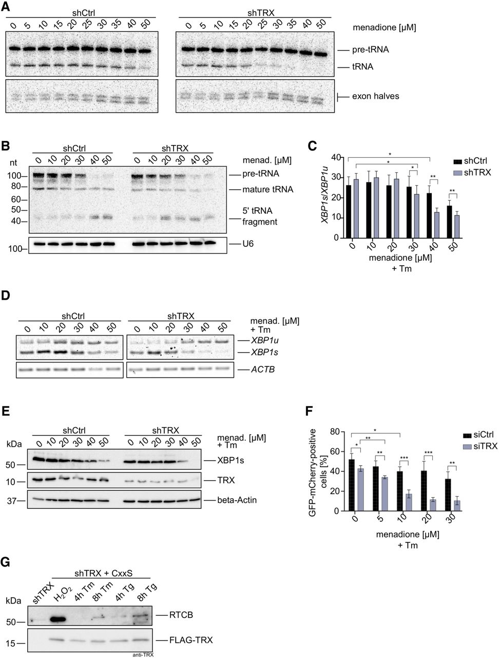
Depletion of TRX impairs the physiological functions of the tRNA-LC. (A) ShCtrl and shTRX cells were treated with indicated menadione concentrations for 1 h before harvest and lysis. An internally radiolabeled S. cerevisiae pre-tRNA substrate was added to cell lysates to monitor cleavage by the TSEN complex and ligation to mature tRNAs by the tRNA-LC. Pre-tRNA processing was monitored by denaturing urea gel electrophoresis and visualized by phosphorimaging. (B) ShCtrl and shTRX were treated with menadione for 1 h. RNA was isolated and subjected to northern blot analysis using a probe complementary to the 5′exon of Tyr-tRNA. A probe targeting U6 snRNA was used as a loading control. (C) ShCtrl and shTRX cells were treated with indicated menadione concentrations and 1.5 µg/mL Tm for 4 h before RNA isolation and cDNA synthesis. RT-qPCR was performed to measure XBP1s and XBP1u mRNA levels relative to ACTB (shown as the ratio XBP1s/XBP1u). (n = 9, mean values ± SEM are shown. Significances were analyzed using paired Student's t-test assuming unequal variances, with [*] P < 0.05; [**] P < 0.01; [***] P < 0.005). (D) ShCtrl and shTRX cells were treated with indicated menadione concentrations and 1.5 µg/mL Tm for 4 h before RNA isolation and cDNA synthesis. Semiquantitative RT-PCR was performed to analyze levels of XBP1 mRNA and ACTB mRNA. PCR products were resolved by agarose gel electrophoresis. (E) ShCtrl and shTRX cells were treated with indicated menadione concentrations and 1.5 µg/mL Tm for 4 h before harvest. Cells were lysed and protein levels of XBP1s and TRX were analyzed by western blot using respective antibodies. β-Actin levels were assessed as a loading control. (F) HEK293 FITR cells expressing GFP-XBP1intron-mCherry were transfected with siRNA pools targeting TXN (siTRX) or control siRNA (siCtrl) for 3 d. Expression of the reporter was induced for 5 h with 2 µg/mL Dox and treatment with menadione, and 1.5 µg/mL Tm was performed for 4 h. Splicing of the intron (generation of GFP-mCherry fusion protein) was assessed by FACS analysis. The graph only shows GFP-mCherry positive cells (n = 4, mean values ± SEM are shown. Significances were analyzed using unpaired Student's t-test assuming unequal variances, with [*] P < 0.05; [**] P < 0.01; [***] P < 0.005). (G) ShTRX FLAG-TRX CxxS cells were treated for 4 or 8 h with 1.5 µg/mL Tm, or for 4 or 8 h with 300 nM Tg before lysis. As a positive control for trapping, shTRX FLAG-TRX CxxS cells were treated for 3 min with 100 µM H2O2. ShTRX cells were left untreated as a negative control. Immunoprecipitation and elution were performed in nonreducing conditions. Eluates were analyzed by western blot using anti-RTCB or anti-TRX antibodies.




