


Hammerhead ribozyme-based U-insertion and deletion RNA editing assays for multiplexing in HTS applications
- 1Institute of Parasitology, McGill University, Ste. Anne de Bellevue, Quebec, Canada H9X 3V9
- 2Department of Biochemistry, McGill University, Montreal, Quebec, Canada H3G 1Y6
- Corresponding author: reza.salavati{at}mcgill.ca
Abstract
Untranslatable mitochondrial transcripts in kinetoplastids are decrypted post-transcriptionally through an RNA editing process that entails uridine insertion/deletion. This unique stepwise process is mediated by the editosome, a multiprotein complex that is a validated drug target of considerable interest in addressing the unmet medical needs for kinetoplastid diseases. With that objective, several in vitro RNA editing assays have been developed, albeit with limited success in discovering potent inhibitors. This manuscript describes the development of three hammerhead ribozyme (HHR) FRET reporter-based RNA editing assays for precleaved deletion, insertion, and ligation assays that bypass the rate-limiting endonucleolytic cleavage step, providing information on U-deletion, U-insertion, and ligation activities. These assays exhibit higher editing efficiencies in shorter incubation times while requiring significantly less purified editosome and 10,000-fold less ATP than the previously published full round of in vitro RNA editing assay. Moreover, modifications in the reporter ribozyme sequence enable the feasibility of multiplexing a ribozyme-based insertion/deletion editing (RIDE) assay that simultaneously surveils U-insertion and deletion editing suitable for HTS. These assays can be used to find novel chemical compounds with chemotherapeutic applications or as probes for studying the editosome machinery.
Keywords
INTRODUCTION
Trypanosoma brucei subsp., Trypanosoma cruzi, and the Leishmania spp. are parasitic trypanosomes that cause devastating endemic diseases such as human African trypanosomiasis (HAT), Chagas disease, and leishmaniases, respectively (Stuart et al. 2008; WHO 2015). Treatments available against these diseases are not ideal due to toxicity, inefficacy, and the emergence of resistant parasite strains, while vaccine development remains a challenge (Denise and Barrett 2001; Fairlamb 2003; Delespaux and de Koning 2007; Field et al. 2017; Pramanik et al. 2019); hence, there is a pressing need for developing novel trypanocidal therapeutics. Among several unusual biochemical features of the trypanosomes, mitochondrial RNA editing is of considerable interest for drug discovery and development (Fidalgo and Gille 2011; Salavati et al. 2012; Field et al. 2017). Most mitochondrial transcripts in these organisms are encrypted and require extensive post-transcriptional modifications through specific insertions and/or deletions of uridylate (U) residues in a guide RNA-dependent manner for the maturation (Stuart et al. 2005; Salavati et al. 2012), ultimately coding for multiple protein components in the oxidative phosphorylation system (Hajduk and Ochsenreiter 2010; Goringer 2012).
An ∼800 kDa multiprotein RNA-editing catalytic complex (RECC) catalyzes the trypanosomatid U-indel RNA editing as dictated by short complementary guide RNA (gRNA) (Rusche et al. 1997; Aphasizhev et al. 2003; Panigrahi et al. 2003; Golas et al. 2009; Li et al. 2009; Aphasizhev and Aphasizheva 2011; Goringer 2012; Voigt et al. 2018; Aphasizheva et al. 2020). Editing catalysis is coordinated in an enzymatic cascade commencing upon hybridization of the 5′ anchor region of a guide RNA to its cognate premature mRNA just downstream from the first editing site. Catalysis initiates with endonucleolytic cleavage at the editing site by an RNA editing endonuclease, followed by U-insertion with a terminal uridylyl transferase (TUTase) or U-deletion with a U-specific exoribonuclease (ExoUase), and terminates with ligation of the edited site with an RNA editing ligase (KREL) (Blum et al. 1990; Pollard et al. 1992; Piller et al. 1995; Corell et al. 1996; Voigt et al. 2018; for a recent review, see Aphasizheva et al. 2020). The edited site then serves as the anchor region for the following gRNA; numerous accessory proteins and protein complexes such as the RNA editing substrate complex (RESC) govern and ensure RNA chaperoning, editing initiation, progression, efficiency and fidelity (Read et al. 2016). The RECC, RESC, and RNA-editing helicase REH2 (REH2C) complexes constitute the functional editosome holoenzyme (Aphasizheva et al. 2014, 2020). The mechanisms through which these protein complexes assemble, interact, and process RNA substrates, are yet to be elucidated. Discovering novel RNA editing inhibitors will serve as excellent tools for unraveling this intricate biochemical process and provide potential chemical scaffolds for therapeutic needs (Mehta et al. 2020).
Based on the traditional editing assays that use radiolabeled RNA substrates (Kable et al. 1996; Seiwert et al. 1996; Rusche et al. 1997; Igo et al. 2000, 2002; Carnes et al. 2005), several suitable high-throughput screening (HTS) in vitro assays have been developed over the past decade. An electrochemiluminescent aptamer-based “full-round” insertion editing assay developed by Liang and Connell (2009), generates a signal upon binding of the conformationally changed aptamer, upon successful editing, to streptavidin-coated microtiter wells (Liang and Connell 2009). A HHR reporter-based “full-round” deletion editing assay designed by Moshiri and Salavati (2010), generates an active ribozyme from successful editing, which cleaves a FRET substrate for an indirect but amplified quantification of the RNA editing (Moshiri and Salavati 2010). Zimmermann et al. (2016) devised a FRET-based RNA ligation assay that uses recombinant RNA editing ligase 1 (KREL1) with a truncated version of recombinant kinetoplastid RNA editing protein A2 (KREPA2). When the above three assays were screened against the library of pharmacologically active compounds (LOPAC1280; Sigma), a different set of potential hits was identified (Liang and Connell 2010; Moshiri et al. 2015; Zimmermann et al. 2016). While common inhibitors of RNA editing were anticipated with similar mechanisms of action, at least against the RNA ligation activity, no such overlap in the hits and their mechanisms were corroborated upon further investigation. Contradicting results may stem from differences in assay sensitivity, variance in source protein composition, chemical substrate concentrations (such as ATP), and incomprehensive mechanism of action (MOA) studies (Table 1).
Summary of RNA editing assays amenable to HTS
Recently, Del Campo et al. (2020) introduced a novel fluorescence-based insertion/deletion editing assay that utilizes capillary electrophoresis to provide quantitative information on editing products and intermediates. While the appealing nature of this assay is its ability to monitor not just the final edited product but also several editing intermediates in a high-throughput manner, high chemical substrate concentrations (0.5 mM ATP and 0.1 mM UTP) could potentially thwart the chances of finding competitive inhibitors of the catalytic enzymes.
This manuscript demonstrates the suitability of HHR-based “precleaved” (PC) RNA editing assays for use in HTS applications or as secondary assays that can aid in determining the MOA of known RNA editing inhibitors. PC insertion and deletion assays are significantly more efficient than a “full-round” editing assay, bypassing the rate-limiting endonucleolytic cleavage step. Furthermore, multiplexed monitoring of U- insertion and deletion, simultaneously, is feasible with the PC ribozyme-based insertion/deletion editing (RIDE) assay, paving the way for more efficient inhibitors of the editosome.
RESULTS
Precleaved RNA editing assay development
To monitor the guide RNA-dependent enzymatic activity of the editosome proteins, we developed three PC HHR-based in vitro assays (namely PC-ligation, PC-deletion, and PC-insertion) coupled with a FRET reporter system (Fig. 1). We trimmed and modified the original version of preedited hammerhead A6Rbz (Wang et al. 2002; Moshiri and Salavati 2010; Moshiri et al. 2015) to bypass the rate-limiting endonucleolytic step by using two fragments of the preedited RNA to mimic the editing site post-cleavage, hence termed “precleaved.” We used gHHR as the guide RNA in PC-insertion assay and its modified version gHHRc (gHHR with an additional cytidine) for PC-ligation and PC-deletion assays. Inclusion of cytidine in the guide RNA was shown to improve in vitro editing efficiency (Supplemental Fig. S2) and was previously described (Cruz-Reyes et al. 2001). As depicted in Figure 1, PC-deletion and PC-insertion assays comprise two consecutive enzymatic steps: uridine deletion or insertion dictated by the mismatches against the template gRNA sequence, catalyzed respectively by an ExoUase or a TUTase before ligation. The ligation assay only requires the final ligation step mediated by a KREL to obtain the final active HHR (Supplemental Fig. S3). The edited product is then detached from the gRNA via adding a gRNA competitor in molar excess. Subsequently, the edited active HHR enzymatically cleaves a FRET-labeled substrate RNA, measured by the signal detected that corresponds to the amount of edited product. Upon testing with intermediate HHR sequences that could potentially arise from these assays, only the fully edited HHR was observed to cleave its FRET substrate (Supplemental Fig. S4).
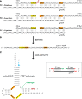
Schematic representation of the precleaved RNA editing assays. (A) Trimolecular hybrids are involved in the PC-deletion, -insertion, and -ligation assays. Three U residues were designed for removal from the 5′Del fragment in PC-deletion; one missing U residue was designed for addition to the 3′Ins fragment in PC-insertion; no requirement for U-deletion or -insertion in PC-ligation. The Watson–Crick and G·U wobble base pairs are depicted by solid and dashed lines. The critical nucleotides of the catalytic site of the HHR are indicated in bold and highlighted in yellow and orange (B) RNA editing results in the formation of an active HHR. (C) Enzymatic cleavage activity of the active HHR on the FRET substrate (containing a fluorophore and a quencher). An arrow indicates the cleavage site. The inset depicts the usage of nonconventional termini in the RNA substrates to reduce/eliminate aberrant editing products. Guide RNA gHHRc (gHHR with cytidine in the editing site) was used for efficient RNA editing (Supplemental Fig. S2).
The PC-deletion and insertion in vitro RNA editing assays were initially monitored using a radiolabeled version of the 5′ HHR fragment (Fig. 2A,B, upper panels). In these experiments, we included three control conditions. In the absence of editosome proteins, there was no change to the radiolabeled 5′ HHR fragment (condition 1). Furthermore, in the absence of gRNA, there was nonspecific removal of nucleotides in PC deletion and, intriguingly, correct addition of a uridine residue in PC insertion (condition 2). The absence of the 3′HHR fragment resulted in correct removal of uridine residues likely dictated by the gRNA (condition 3). Other control conditions containing known editing inhibitors (Amaro et al. 2008; Durrant et al. 2010; Liang and Connell 2010; Moshiri et al. 2011; Salavati et al. 2012; Zimmermann et al. 2016; Mehta et al. 2020), such as the sulfonated compounds mordant black 25 (MrB) (condition 4) and suramin (condition 5), completely inhibited RNA editing activity. The conditions assayed were replicated in the FRET-based versions for comparison, which resulted in a quantifiable signal obtained only from the fully edited HHR (Fig. 2A,B, lower panels). Minuscule editing activity was observed without added ATP (condition 6), likely due to the purified editosome containing preadenylated KREL proteins. While robust editing was detected in the presence of added ATP in the radiolabeled assay on urea PAGE (condition 7), it surprisingly did not translate to a correlated amount of HHR activity. Upon closer investigation of the final product on a 6% urea PAGE, the final edited product did not correspond to the same length as the positive control active HHR (Supplemental Fig. S5), indicating that ligation of the 2 HHR fragments occurs rather prematurely. This was circumvented by the delayed addition of ATP 1-h post incubation initiation (condition 8 in Fig. 2; Supplemental Fig. S5).
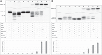
Development of precleaved deletion and insertion assays. Assays are monitored using radiolabeled HHR RNA substrates (top panel) and FRET-based HHR substrates (bottom panel) with several controls (as described in the middle panel). (A) PC-deletion assay and (B) PC-insertion assay with all components except functional editosome (lane 1), guide RNA (lane 2) and 3′ fragment (lane 3). Both assays in the presence of RNA editing inhibitors (MrB and suramin, lanes 4 and 5). A faint product/signal was detected without ATP (lane 6). In the presence of all reaction components with discernible product/signal (lane 7). Improved HHR activity is observed when ATP addition is delayed by 1-h post reaction initiation (lane 8). Active HHR (0.5 and 1.5 pmol) was used as a size and activity control in the PC-insertion and deletion assay, respectively, to magnify the enhancement in activity (lane 9). Means and standard deviations in the bottom plot were obtained from four replicates. Lanes 6,7, and 8 in both panels were significantly different from no protein controls (lane 1) (P < 0.05) by one-way ANOVA (GraphPad Prism).
Optimizing the precleaved assay conditions
The FRET-based PC insertion and deletion assays were screened to establish the optimized concentrations of ATP, UTP (for PC insertion and the RIDE assay), and purified editosome (KREL1-TAP tag calmodulin eluate) and also for optimized incubation durations (Fig. 3). The optimized ATP concentration was established approximately at 100 nM in the PC deletion assay (Fig. 3A) and PC insertion assay (data not shown), without compromising significantly on efficiency. Likewise, the optimized UTP concentration for the PC insertion assay was observed approximately at 10 µM (Fig. 3B). A linear correlation between the amount of editosome and relative editing activity in the PC assays was observed (Fig. 3C). The optimized ATP (100 nM) and UTP (10 µM) concentration with 50 fmol of editosome were used in the timepoint assay (Fig. 3D), also exhibiting a linear relationship between time and activity, whereby overnight incubation led to the most substantial activity for all assays. Note that the timepoint initiation was after addition of ATP. The output of the optimized assays, as compared in Figure 3E against a standard curve of active HHR activity, indicates efficient activities from PC deletion and ligation.
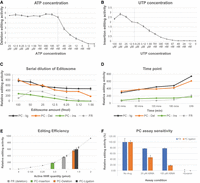
Optimized conditions for efficient editing in the precleaved assays. Varying concentrations of (A) ATP and (B) UTP substrates in the PC-deletion and insertion assays, respectively. (C) Relative RNA editing activities of different amounts of functional editosome in the PC-insertion, deletion, ligation, and the full-round assay. (D) Timepoint measurement of editing activities in the PC-insertion, deletion, ligation, and the conventional full-round assay. (E) Editing efficiencies of the different assays against a standard plot of active HHR. Each bar represents an editing assay and the corresponding activity of their edited product. (F) an ATP analog (α, β-methylene ATP) was used to compare the sensitivity of the ligation assay with a full-round assay to find competitive inhibitors. There was no difference between editing activity in the full round assay in the presence of 20 µM versus 100 µM ABMA (P > 0.05), but the ligation assay inhibition was significant (P < 0.05), calculated by one-way ANOVA (GraphPad Prism). Means and standard deviations in all six plots were calculated and are shown for at least two replicates.
As the PC assays use substrate ATP at 100 nM, 10,000-fold less than the conventional full-round ribozyme-based FRET assay, these assays are more sensitive to discovering competitive inhibitors of ATP. In Figure 3F, inhibition of the PC ligation assay by a nonhydrolyzable ATP analog, α, β-methylene ATP (ABMA), is observed. However, no such impact is seen on the “full-round” assay. The amenability of these assays in HTS applications was then determined through Z-factor calculation with at least 20 replicates in the presence and absence of a known inhibitor (suramin). The Z-factor determined for the PC ligation, deletion and insertion assays were 0.7, 0.82, and 0.84, respectively (data not shown).
Ribozyme insertion/deletion editing (RIDE) assay
To monitor insertion and deletion editing activities simultaneously, the HHR sequence was slightly modified to recognize a different FRET substrate (Fig. 4A). When tested in the same reaction, the active HHR1 and HHR2 reporters specifically cleave FRET1 (measured for FAM) and FRET2 (measured for Cy5) substrates, respectively (Fig. 4B). PC versions of these HHR substrates were thus designed to facilitate multiplexing U-insertion, and deletion editing activities in the RIDE assay (Fig. 4C), where HHR1 is modified for use in U-insertion editing and HHR2 is modified for screening U-deletion editing. Figure 4D shows the edited products of U-insertion and U-deletion reactions to cleave their respective FRET1 and FRET2 substrates specifically. Consequently, multiplexing is feasible by combining these assays, where FRET signals representing both U-insertion and U-deletion activities are detected simultaneously, as corroborated on urea-PAGE (Supplemental Fig. S6). Intriguingly, in the absence of UTP, a small amount of FRET1 cleavage (from the U-insertion HHR reporter) is observed, likely due to recycling of uridine residues obtained from the U-deletion HHR substrate catalysis.
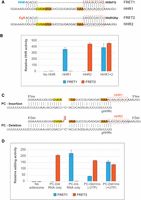
Multiplex measurement of U-insertion and deletion in the ribozyme-based insertion/deletion editing (RIDE) assay. (A) Schematic representation of HHR1 and HHR2 bound to their respective target FRET substrates, FAM-FRET1- IABkFQ and Cy5-FRET2- IAbRQSp. The dissimilar FRET substrate hybridizing region of the two HHRs is shown in red dashed boxes. Essential nucleotides in the catalytic site of the ribozyme are highlighted in yellow and orange. (B) Specificity of HHR1 and HHR2 against their FRET substrates. Signals released because of FRET1 and FRET2 cleavage are shown in blue (FAM) and orange (Cy5) for four experimental conditions. All four lanes contain both FRET1 and FRET2 substrates. The x-axis determines which ribozyme is added to each lane. (C) Diagram of PC-insertion and PC-deletion trimolecular hybrids in the RIDE assay. HHR1 and HHR2 were fragmented to provide the PC RNA substrates for assaying insertion and deletion activities. (D) Measurement of U-insertion and -deletion activities from the RIDE assay. FRET1 and FRET2 substrates are added post-editing. Means and standard deviations were obtained from three replicates, and the values corresponding to each colored bar were statistically significant (P-value < 0.05) by one-way ANOVA (GraphPad Prism).
DISCUSSION
Trypanosomatid RNA editing is suitable for target-based drug discovery and development (Salavati et al. 2012). While designing efficacious RNA editing assays is key to finding novel inhibitors, implementing an 800 kDa multiprotein complex in HTS has previously led to discovering nonspecific inhibitors. Apart from the recent finding that the editosome complex is prone to nonspecific inhibition by negatively charged compounds, likely due to its reliance on the positively charged MRP1/2 proteins for RNA chaperoning and editing initiation (Mehta et al. 2020), the assays presented in this paper have been optimized to overcome the other shortcomings with the assays currently in use. Bypassing the rate-limiting endonucleolytic cleavage step of RNA editing (Igo et al. 2000, 2002; Carnes and Stuart 2007) with the help of PC RNA substrates, not only improves editing efficiency as observed in Figure 3E, but also lowers the requirement of substrate ATP by 100- to 10,000-fold (Table 1), enabling heightened sensitivity of these assays in discovering ATP competitive inhibitors (Fig. 3F). Moreover, with the development of the multiplexing RIDE assay, simultaneous measurements of TUTase, ExoUase and ligase activities further increase the odds of discovering inhibitors of these catalytic processes. Upon multiplexing the sensitivity for finding competitive inhibitors such as ABMA was maintained (Supplemental Fig. S7).
During optimization of these assays, two factors played a role: (i) inclusion of a cytidine residue in the gRNA sequence and (ii) delayed addition of ATP. The addition of a cytidine residue in the RNA editing site region of the gRNA, significantly enhances U-deletion and ligation activity (Supplemental Fig. S2), as previously described (Cruz-Reyes et al. 2001; Wang et al. 2002). As it does not affect the efficiency of the PC insertion assay (Supplemental Fig. S2), either gRNA could be used for that assay, gHHR or gHHRc; therefore, to limit compromising the U-deletion efficiency in the RIDE assay, gHHRc is used in both trimolecular hybrids (Fig. 4A). The requirement of delayed addition of ATP stems from the observation in Figure 2 condition 7, where the ligated products do not appear to be completely edited on urea-PAGE (further resolved on a 6% urea-PAGE in Supplemental Fig. S5). This indicates that the ligases are more efficient than the other enzymes in the RECC, which likely already contain preadenylated ligases (Fig. 2, condition 6). This could presumably be why there is inefficiency in the “full-round” assays, where the edited site is immediately ligated post-endonucleolytic cleavage. To circumvent this issue, the assay is designed to initiate in the absence of ATP for 1 h to allow complete insertion or deletion of uridine residues as per the gRNA sequence (Supplemental Fig. S5), and significantly increase the activity of the final edited HHR (Fig. 2, condition 8).
The assays presented exhibit a linear relationship with the amount of purified editosome, and robust activity is obtained from 1 h post incubation (1 h following ATP addition). Moreover, the RNA substrates do not require extensive chemical modifications to impart stability. Monitoring FRET substrate can be performed in real-time with kinetic readings on a quantitative RT-PCR machine or performed endpoint after incubation for 30–60 min with the FRET substrate, depending on the instrumentation available for HTS. Furthermore, preliminary testing establishes the feasibility of these assays for use in HTS applications with reconstituted catalytic editosomes with recombinant proteins, as performed earlier (Kang et al. 2005, 2006). The major benefit of using reconstituted editosome is to obtain inhibitors only against the proteins involved, rather than obtaining nonspecific inhibitors as previously observed, such as the sulfonated compounds that target the MRP1/2 proteins (Moshiri et al. 2011; Mehta et al. 2020). In conclusion, this manuscript presents novel PC assays that are amenable for multiplexing in HTS applications and can also serve as secondary assays that are vital in determining the mechanism of known RNA editing inhibitors.
MATERIALS AND METHODS
Preparation of RNA substrates
The PC RNA substrates used in this study (as summarized in Table 2) were designed based on the ribozyme reporter previously described (Moshiri and Salavati 2010). In this sequence, additional U residues in the HHR active site served as substrates for the deletion assay, while the removal of a U residue from the HHR active site served as substrates for the insertion assay. For the ligation assay, no modifications to the HHR active site were required (Fig. 1). These substrates were synthesized and HPLC purified by Integrated DNA Technologies (IDT). To minimize any aberrant modifications to the RNA substrates, the 5′ ends of the 5′ HHR fragments and gRNA were hydroxylated, and the 3′ends of the 3′ HHR fragments and gRNA were phosphorylated (Fig. 1, inset).
RNA substrates used in the precleaved assays
For use in radiolabeled editing assays, as performed conventionally on urea denaturing polyacrylamide gel electrophoreses (PAGE), the 5′Del and 5′Ins fragments were 5′ end labeled with [γ-32P] ATP using T4 PNK (New England Biolabs) and subsequently purified using 8% urea PAGE as described before (Wang et al. 2002).
Purification of Trypanosoma brucei editosome
Genetically modified procyclic T. brucei expressing TAP-tagged kinetoplastid RNA editing ligase 1 (KREL1) was used for functional editosome purifications as described before (Stuart et al. 2004). Antibodies against four RECC proteins (KREL1, KREPA1, KREPA2, and KREPA3) were used for probing the purified elutions by western blotting (Supplemental Fig. S1).
Precleaved assays: development and optimization
Both versions of editing assays, FRET-based and radiolabeled, contained 2 pmol of the 5′ and 3′ HHR fragments, and 4 pmol of gRNA for the respective type of editing (refer to Table 2; Fig. 1). Before starting the assays, the RNA mixtures were hybridized by denaturation at 70°C for 5 min in a water bath and allowed to slow-cool to ambient room temperature, typically lasting 40–60 min. Following hybridization, the RNA mixture was added to a master mix to obtain a final reaction composition of 25 mM HEPES (pH 7.9), 10 mM Mg (OAc)2, 50 mM KCl, 1 mM EDTA, 0.1 µM adenosine triphosphate (ATP), 5 mM CaCl2 and 0.1% Triton X-100. The solution also contained 1 µL of the purified editosome (calmodulin eluate from KREL1-TAP tag purification, at ∼40 ng/mL) and 10 µM UTP for insertion editing or RIDE assays. Separate master mixes were prepared for the controls, typically missing one or more reaction components or containing known RNA editing inhibitors (Fig. 2). The plates were sealed and incubated at 28°C overnight (∼16–20 h).
To stop the editing reaction in the radiolabeled assays, 40 pmol of an appropriate DNA guide competitor was added to each reaction, along with 0.1 volumes of 3 M sodium acetate. The RNA was then extracted with phenol:chloroform:isoamyl alcohol (25:24:1) and precipitated with ethanol before reconstitution with a urea loading dye and loading onto a 15% or 6% urea PAGE (30–40 cm gel with S2 sequencing gel electrophoresis apparatus). After running the PAGE for approximately 1.5–2 h at 45–50 watts, the gel was scanned on a PhosphorImager (Bio-Rad).
To stop the editing reaction in the FRET-based assays, 40 pmol of an appropriate DNA guide competitor was added to each well and incubated at 85°C for 10 min. After cooling at RT for 5 min, 20 pmol of the HHR FRET substrate were added to each well and read kinetically for fluorescein every min at 37°C in an RT-qPCR machine. The slope calculated from the kinetically measured fluorescent signal outputs represents editing activity. Alternatively, the final measurement could be taken as an endpoint reading after 30–60 min incubation at 37°C. Assay optimization was facilitated by measuring various ATP, UTP, and purified editosome concentrations while testing for different incubation durations.
Ribozyme insertion/deletion editing (RIDE) assay
Multiplexing in the RIDE assay was performed with minor modifications to the HHR sequence for PC deletion, requiring a different 3′ fragment and HHR FRET substrate (Table 2; Figs. 1, 4). The editing reaction was performed as above, with two trimolecular hybrids ([5′Del, 3′Del2, gHHRc] and [5′Ins, 3′Ins, gHHRc]) prepared independently before assay initiation. For signal measurement, 20 pmol of both HHR substrates, FRET1 and FRET2, were added to the reaction simultaneously to read for fluorescence from the FAM and Cy5 fluorophores.
Z′ factor determination
The Z′ was calculated for the assays using the following formula where σ is the standard deviation and µ is the mean of the
values (Zhang et al. 1999). n and p stand for negative and positive controls. The number of replicates for each control was 10 and suramin was used
as the inhibitor of the RNA editing reaction.

SUPPLEMENTAL MATERIAL
Supplemental material is available for this article.
ACKNOWLEDGMENTS
We thank Arezou Kamelshahroudi and Akshaya Srikanth for their technical assistance. This work was supported by the National Institutes of Health grant R01AI143593 to R.S.
Footnotes
-
Article is online at http://www.rnajournal.org/cgi/doi/10.1261/rna.079454.122.
-
Freely available online through the RNA Open Access option.
- Received September 29, 2022.
- Accepted November 18, 2022.
This article, published in RNA, is available undera Creative Commons License (Attribution-NonCommercial 4.0 International), as described at http://creativecommons.org/licenses/by-nc/4.0/.
REFERENCES
MEET THE FIRST AUTHOR

Meet the First Author(s) is an editorial feature within RNA, in which the first author(s) of research-based papers in each issue have the opportunity to introduce themselves and their work to readers of RNA and the RNA research community. Mojtaba Rostamighadi is the first author of this paper, “Hammerhead ribozyme-based U-insertion and deletion RNA editing assays for multiplexing in HTS applications.” Mojtaba is a PhD candidate in Dr. Reza Salavati's laboratory at the institute of parasitology at McGill University. His laboratory studies kinetoplastid parasites with an emphasis on RNA editing, seeking novel treatments, using target-based drug discovery, for the diseases they cause.
What are the major results described in your paper and how do they impact this branch of the field?
We developed an efficient fluorescent-based multiplex assay that can monitor the activity of multiple enzymes of the editosome simultaneously, offering more chances of finding new inhibitors. Also, low concentrations of enzyme substrates such as ATP and UTP enable our assay to find competitive inhibitors. This new assay is high-throughput amenable and can be used to screen large libraries of compounds.
What led you to study RNA or this aspect of RNA science?
The unique type of RNA editing in kinetoplastids and the massive protein complex mediating this pathway made me wonder how these parasites developed such a complex pathway. Even after decades, we have yet to learn about the dynamics of the editosome.
If you were able to give one piece of advice to your younger self, what would that be?
Stay focused and learn more every day, no matter what!
What are your subsequent near- or long-term career plans?
I want to establish a start-up company to find treatments for parasitic diseases. Doing what you like to do always makes you a successful person.




