


A selective and sensitive detection system for 4-thiouridine modification in RNA
- 1Department of Materials Science and Biotechnology, Graduate School of Science and Engineering, Ehime University, Matsuyama, Ehime 790-8577, Japan
- 2Cellular and Molecular Biotechnology Research Institute, National Institute of Advanced Industrial Science and Technology (AIST), Koto-ku, Tokyo 135-0064, Japan
- Corresponding author: hori.hiroyuki.my{at}ehime-u.ac.jp
Abstract
4-Thiouridine (s4U) is a modified nucleoside, found at positions 8 and 9 in tRNA from eubacteria and archaea. Studies of the biosynthetic pathway and physiological role of s4U in tRNA are ongoing in the tRNA modification field. s4U has also recently been utilized as a biotechnological tool for analysis of RNAs. Therefore, a selective and sensitive system for the detection of s4U is essential for progress in the fields of RNA technologies and tRNA modification. Here, we report the use of biotin-coupled 2-aminoethyl-methanethiosulfonate (MTSEA biotin-XX) for labeling of s4U and demonstrate that the system is sensitive and quantitative. This technique can be used without denaturation; however, addition of a denaturation step improves the limit of detection. Thermus thermophilus tRNAs, which abundantly contain 5-methyl-2-thiouridine, were tested to investigate the selectivity of the MTSEA biotin-XX s4U detection system. The system did not react with 5-methyl-2-thiouridine in tRNAs from a T. thermophilus tRNA 4-thiouridine synthetase (thiI) gene deletion strain. Thus, the most useful advantage of the MTSEA biotin-XX s4U detection system is that MTSEA biotin-XX reacts only with s4U and not with other sulfur-containing modified nucleosides such as s2U derivatives in tRNAs. Furthermore, the MTSEA biotin-XX s4U detection system can analyze multiple samples in a short time span. The MTSEA biotin-XX s4U detection system can also be used for the analysis of s4U formation in tRNA. Finally, we demonstrate that the MTSEA biotin-XX system can be used to visualize newly transcribed tRNAs in S. cerevisiae cells.
Keywords
INTRODUCTION
4-Thiouridine (s4U) is a modified nucleoside (Lipsett 1965), found at positions 8 and 9 in tRNA from eubacteria and archaea (Boccaletto et al. 2018; Sajek et al. 2020). The biosynthetic pathways of s4U are different in eubacteria and archaea (Shigi 2014; Čavužić and Liu 2017; Zheng et al. 2017, 2021; Shigi 2021). In Escherichia coli, the sulfur atom in l-cysteine is activated by cysteine desulfrase (IscS) and is then transferred to tRNA by tRNA 4-thiouridine synthetase (ThiI) (Mueller et al. 1998; Kambampati and Lauhon 1999; Lauhon and Kambampati 2000). Two cysteine residues, which form a disulfide bond, in E. coli ThiI are essentially required for the reaction (Palenchar et al. 2000; Mueller et al. 2001; Veerareddygari et al. 2016). However, the iscS gene is not present in the majority of archaea genomes (Liu et al. 2012). In the case of Methanococcus maripaludis, ThiI contains an Fe-S cluster and S2− is used as the sulfur donor instead of l-cysteine (Liu et al. 2012). However, the Fe-S cluster type thiI gene is not present in some archaea genomes; the biosynthetic pathway of s4U in these organisms has not been determined (Tomikawa et al. 2013; Čavužić and Liu 2017).
The physiological roles of s4U in tRNA have been gradually elucidated. The s4U8 modification in tRNA functions as an ultraviolet light (UV)-resistance factor in bacterial cells (Ramabhadran et al. 1976). Irradiation of near-UV causes cross-linking between s4U8 and C13 in tRNA (Favre et al. 1969). Accumulation of cross-linked tRNA in E. coli cells slows down protein synthesis and the cell growth rate (Caldeira de Araujo and Favre 1985). This growth delay induces the DNA repair system (Caldeira de Araujo and Favre 1986). Furthermore, the s4U8 modification contributes to stabilize the L-shaped tRNA structure because the melting temperature of tRNASer from an E. coli thiI gene deletion strain decreases by 4.7°C as compared to that from the wild-type strain (Nomura et al. 2016). Moreover, the s4U8 modification works as a tRNA quality control system in Vibrio cholerae in the stationary growth phase (Kimura and Waldor 2019).
Beyond the natural functions, s4U has been utilized as a biotechnological tool for analysis of RNAs because s4U is less cytotoxic than artificial uridine derivatives and the sulfur atom in s4U is highly reactive (Heiss and Keller 2017; Zheng et al. 2017). For example, pulse labeling of RNA by s4U has been utilized for analysis of newly transcribed RNA and/or RNA degradation (Johnson et al. 1991; Cleary 2008; Miller et al. 2009; Rabani et al. 2011, 2014; Gay et al. 2013; Duffy et al. 2015, 2018; Fuchs et al. 2015; Duffy and Simon 2016; Herzog et al. 2017; Hida et al. 2017; Riml et al. 2017; Russo et al. 2017; Schofield et al. 2018; Gregersen et al. 2020). Furthermore, cross-linking by s4U, which is artificially introduced into a target RNA(s), has been used for the studies of RNA–RNA and RNA–protein interactions (Juzumiene and Wollenzien 2001; Garzia et al. 2017; Soszynska-Jozwiak et al. 2021). Moreover, s4U has been used for the site-directed labeling of RNAs (Hara et al. 1970; Caron and Dugas 1976; Qin et al. 2003; Wunnicke et al. 2011). Therefore, a selective and sensitive s4U detection system is required in RNA technology and tRNA modification fields.
Historically, s4U in tRNA has been detected by UV absorption (Lipsett 1965), circular dichronism (CD) spectrum (Hori et al. 1989; Milder et al. 1989), chemical probing by bromoacetamide derivatives (Hara et al. 1970; Caron and Dugas 1976), cyanoethylation by acrylonitrile (Ofengand 1967), [(N-acryloylamino)phenyl]mercuric chloride (APM)-gel electrophoresis (Igloi 1988), liquid chromatography–mass spectrometry (LC-MS) (Sakaguchi et al. 2015; Ross et al. 2016), and chemical labeling by fluoresceinamine (FAM-NH2) (Watson et al. 1995; Wang et al. 2019). However, measurements by UV absorption and CD require relatively large amounts of RNA. Chemical probing by bromoacetamide derivatives cannot distinguish between s4U and other sulfur-containing nucleosides such as 2-thiouridine (s2U) (Hara et al. 1970; Caron and Dugas 1976). Furthermore, although acrylonitrile cyanoethylation can distinguish s4U and s2U, acrylonitrile reacts with pseudouridine and inosine as well as s4U (Ofengand 1967). An issue with the APM-detection method is that it contains mercury, which is very toxic. LC-MS is reliable; however, an LC-MS system is expensive for general biochemical laboratories and is not suitable for analysis of multiple samples in a short time. Although FAM-NH2 labeling is sensitive, this probe weakly reacts with s2U in addition to s4U (Wang et al. 2019). To overcome these problems, we have devised a selective and sensitive method for detection of s4U in RNA.
RESULTS AND DISCUSSION
Outline of s4U detection system
There are several thiol-specific reagents for labeling of s4U; however, iodoacetamide derivatives react with s2U in addition to s4U (Heiss and Keller 2017). We therefore selected 2-aminoethyl-methanethiosulfonate (MTSEA) for labeling of s4U. MTSEA has been used for fractionation of s4U-labeled mRNA (Rabani et al. 2011; Gregersen et al. 2020) and reactivity (selectivity) of MTSEA toward s2U has not been reported. MTSEA was originally developed as a cysteine-specific chemical modification reagent and has been used in protein chemistry (Rothwarf and Scheraga 1993; Marjamäki et al. 1999). Therefore, biotin-coupled MTSEA (MTSEA biotin-XX: Fig. 1A) is commercially available. If MTSEA biotin-XX can specifically attach to s4U in RNA, bound biotin can be detected by chemiluminescence using streptavidin-horse radish peroxidase (HRP). This system itself was recently reported in the reference (Gregersen et al. 2020). To investigate whether this system is sensitive and quantitative, we initially constructed a model system for s4U detection in RNA (Fig. 1B,C). We prepared two artificial RNAs (Linear-s4U and Linear-U) (Fig. 2A). Linear-s4U contains only one s4U in its sequence while Linear-U contains U instead of s4U. These two RNAs were dissolved in water, sequentially diluted, reacted with MTSEA biotin-XX, recovered by phenol–chloroform treatment and ethanol precipitation, dissolved in water again, spotted onto a Hybond-N+ nylon membrane filter and fixed to the membrane by irradiation with UV at 254 nm. To visualize RNA, the filter was stained with methylene blue. The filter was immersed in streptavidin-HRP solution at 25°C for 90 min. Chemiluminescence derived from bound streptavidin-HRP was monitored. Details are described in the Materials and Methods section.
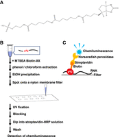
(A) Structure of MTSEA biotin-XX. (B) Outline of MTSEA biotin-XX s4U detection system. RNA containing s4U is reacted with MTSEA biotin-XX and fixed onto a nylon membrane filter. Streptavidin-HRP captures the biotin in the sample. The activity of HRP is detected by chemiluminescence. Details are described in the main text and the Material and Methods section. (C) Schematic drawing of MTSEA biotin-XX s4U detection system.
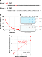
(A) Sequences of Linear-s4U and Linear-U RNAs. Linear-s4U contains s4U only at position 9 from 5′-end. Linear-U contains U instead of s4U. (B) When MTSEA biotin-XX is reacted with s4U, the absorbance at 330 nm derived from s4U disappears. To determine the reaction time, decrease of absorbance at 330 nm in a reaction mixture containing Linear-s4U and MTSEA biotin-XX was monitored. (C) Comparison of the reactivity of Linear-s4U and Linear-U RNAs toward MTSEA biotin-XX. Both RNAs were treated with MTSEA biotin-XX and then fixed onto a nylon membrane filter. RNAs were visualized by methylene blue staining (upper). The bound MTSEA biotin-XX was detected by chemiluminescence (lower). (D) Relationship between the amounts of RNAs and the chemiluminescence intensity of spots showed linearity from 0 pmol to 100 pmol RNAs. Chemiluminescence derived from the Linear-U samples was undetectable. Relative intensity is defined so that the chemiluminescence derived from the 50 pmol Linear-s4U sample is expressed as 1.00.
The s4U detection system is sensitive and quantitative
Linear-s4U and U RNAs were prepared by in vitro T7 RNA polymerase transcription and purified by 10% polyacrylamide gel containing 7 M urea electrophoresis (10% PAGE [7 M urea]). To confirm that Linear-s4U contains s4U instead of U, nucleoside analyses of Linear-s4U and U RNAs were performed. As shown in Supplemental Figure 1, Linear-s4U RNA contains s4U but not U. In contrast, Linear-U contains U instead of s4U. Next, we investigated the reaction time with MTSEA biotin-XX. If s4U in Linear-s4U RNA is reacted with MTSEA biotin-XX, the absorbance at 330 nm derived from s4U disappears. We monitored the decrease in absorbance at 330 nm in the reaction mixture and found that 30 min is sufficiently long reaction time (Fig. 2B). Figure 2C shows the results of experiments with Linear-s4U and Linear-U with a reaction time of 30 min. When Linear-s4U was used (Fig. 2C, upper), chemiluminescence derived from HRP was clearly detected. In contrast, chemiluminescence was not observed with the Linear-U samples (Fig. 2C, lower). This result shows that s4U is detectable by this system. Furthermore, it is also clear that this system can distinguish between s4U and common nucleosides (A, G, C, and U). The chemiluminescence was detectable from a spot of 2.5 pmol Linear-s4U, demonstrating that the sensitivity of the system is comparable or slightly higher than that by the FAM-NH2 labeling method (Wang et al. 2019). To show the detection limit clearly, chemiluminescence from 2.5 pmol of Linear-s4U and -U RNAs were compared rigorously (Supplemental Fig. 2). Furthermore, the chemiluminescence intensity showed linearity up to 100 pmol (Fig. 2D). Taking these experimental results together, we conclude that the MTSEA biotin-XX s4U detection system is sensitive and quantitative.
The MTSEA biotin-XX system can detect s4U in a stem structure
4-Thiouracil can form a base pair with adenine and guanine. In studies of RNA metabolism and RNA structure, target RNA frequently forms a stem–loop structure(s) and s4U is often embedded in the stem. Therefore, chemical probing often requires denaturation of target RNA. To investigate whether the MTSEA biotin-XX system can detect s4U in a stem structure, we prepared RNA with such a structure (Stem-s4U RNA) (Fig. 3A). This RNA was annealed in the presence of 5 mM Mg2+ before use. As shown in Figure 3B and C, the MTSEA biotin-XX system detects s4U in the annealed Stem-s4U: the limit of detection was 10 pmol. To show the detection limit, chemiluminescence from 10 pmol annealed Stem-s4U and -U RNAs were compared (Supplemental Fig. 3). Because MTSEA biotin-XX possesses a long linker region composed of three aminohexyl groups (Fig. 1A), the methanethiosulfonate in MTSEA biotin-XX and s4U in the stem structure seem to react without severe steric hindrance. This experimental result demonstrates that the MTSEA biotin-XX system can detect s4U in a stem structure without denaturation. This is a clear advantage of the MTSEA biotin-XX s4U detection system. Moreover, when the stem structure in Stem-s4U RNA was disrupted by heat and rapid cooling in the presence of 10 mM EDTA, chemiluminescence was observed from a spot of 2.5 pmol Stem-s4U RNA (Fig. 3B,C). Thus, although the MTSEA biotin-XX system can be used without denaturation of target RNA, denaturation improves the limit of detection.
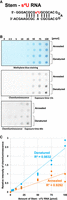
(A) Sequence of Stem-s4U RNA. When this RNA is annealed, s4U is embedded in the stem structure. (B) Annealed and denatured Stem-s4U RNAs were spotted onto a nylon membrane filter and then visualized by methylene blue staining (upper). The chemiluminescence derived from the activity of HRP was monitored (middle and lower). To investigate the detection limit, exposure time was changed from 13 sec (middle) to 60 sec (lower). (C) Relationship between the amounts of RNAs and the chemiluminescence intensity of spots showed linearity. The chemiluminescence intensity of denatured Stem-s4U RNA samples is comparable with that of Linear-s4U RNA samples (Fig. 2D). Relative intensity is defined so that the chemiluminescence derived from the 50 pmol Stem-s4U sample is expressed as 1.00.
MTSEA biotin-XX reacts only with s4U but does not react with other sulfur-containing modified nucleosides in tRNA
When newly transcribed RNAs are visualized by incorporation of s4U, cross reactivity with other sulfur-containing modified nucleosides is an important problem. Sulfur-containing nucleosides exist mainly in tRNAs (Shigi 2014, 2021; Čavužić and Liu 2017; Zheng et al. 2017, 2021). Transfer RNAs from Thermus thermophilus, an extreme-thermophilic eubacterium, abundantly contain sulfur-containing nucleosides, because U54 of all tRNA species in this organism is modified to 5-methyl-2-thiouridine (m5s2U) (Watanabe et al. 1976b; Shigi et al. 2002; Hori et al. 2018; Hori 2019). Although the modification levels of m5s2U54 are different in tRNA species (Takuma et al. 2015), the average of modification levels in all tRNA species is 50% when the cells are cultured at 65°C (Watanabe et al. 1976b; Hori 2019). Furthermore, it has been reported that s4U is contained in T. thermophilus tRNAs (Watanabe et al. 1979; Grawunder et al. 1992; Keith et al. 1993; Tomikawa et al. 2010). In fact, thiI gene is encoded in the T. thermophilus genome (Shigi et al. 2006). T. thermophilus tRNA contains other sulfur-containing nucleosides in addition to s4U and m5s2U because the genes for the corresponding modification enzymes are present in the T. thermophilus genome (Henne et al. 2004; Shigi et al. 2020). For example, MnmA, for 2-thiolation of 5-methylaminomethyl-2-thiouridine derivatives at position 34 in tRNA is encoded in the genome and its enzymatic activity was confirmed (Kambampati and Lauhon 2003; Moukadiri et al. 2014; Shigi et al. 2020). Furthermore, the presence of 2-methylthio-N6-isopentenyladenosine at position 37 in T. thermophilus tRNAPhe has been experimentally demonstrated (Grawunder et al. 1992; Tomikawa et al. 2010). We prepared tRNA fractions from T. thermophilus wild-type and thiI gene deletion (ΔthiI) strains. To confirm the existences of s4U and m5s2U in the tRNA fraction from the wild-type strain, the modified nucleosides were analyzed by high performance C18-liquid column chromatography (Fig. 4A). To detect s4U and m5s2U, absorbance at 330 nm was monitored in addition to 254 nm. In the sample from the wild-type strain, a peak of s4U was clearly observed (Fig. 4A left panels). In contrast, this peak was not observed in the sample from the ΔthiI strain (Fig. 4A right panels). The peak corresponding to m5s2U was observed in the samples from both the wild-type and ΔthiI strains. It should be mentioned that the peak areas of s4U and m5s2U in Figure 4A do not represent the molecular ratio between s4U and m5s2U because the absorbance at 330 nm of m5s2U is very small as compared to that of s4U. Therefore, the s4U and m5s2U in these tRNA fractions were also analyzed by CD (Fig. 4B). In the tRNA fraction from the wild-type strain (red in Fig. 4B), positive CD signals derived from s4U (around 340 nm) and m5s2U (around 315 nm) were clearly observed (Watanabe et al. 1976a; Hori et al. 1989). In contrast, in the sample from the ΔthiI strain (blue in Fig. 4B), the signal derived from s4U was not observed. Thus, we confirmed that the tRNA fraction from the ΔthiI strain contained m5s2U but not s4U. The tRNA fractions were reacted with MTSEA biotin-XX and then loaded onto a polyacrylamide gel containing 7 M urea. After electrophoresis, the tRNAs were electroblotted onto a Hybond-N+ nylon membrane and analyzed by streptavidin-HRP. As shown in Figure 4C, chemiluminescence was observed only in tRNAs from the wild-type strain. This result shows that the MTSEA biotin-XX s4U detection system does not react with s2U derivatives under the tested conditions. Furthermore, we tested whether chemiluminescence from excess amounts of s2U derivatives was detected by the MTSEA biotin-XX system. For this purpose, 300 pmol of tRNA mixtures from T. thermophilus wild-type and ΔthiI strains were used (Supplemental Fig. 4). As shown in Supplemental Figure 4, chemiluminescence was not detected from the ΔthiI tRNA mixture. This result shows that the MTSEA biotin-XX system does not react with the s2U derivatives under the practical conditions. Moreover, we tested whether the MTSEA biotin-XX system can detect s4U in Linear-s4U RNA (Fig. 2A) in the presence of an excess amount of T. thermophilus ΔthiI tRNA mixture. Sequentially diluted Linear-s4U RNA (0, 2.5, 5, and 10 pmol) and 250 pmol T. thermophilus ΔthiI tRNA mixture were mixed and then reacted with MTSEA biotin-XX (Supplemental Fig. 5). Although the intensity of chemiluminescence was weakened, the MTSEA biotin-XX system can detect s4U in 2.5 pmol Linear-s4U RNA in the presence of 250 pmol T. thermophilus ΔthiI tRNA mixture. Thus, the MTSEA biotin-XX system can detect s4U-containing RNA in the presence of an excess amount of s4U-noncontaining RNA.
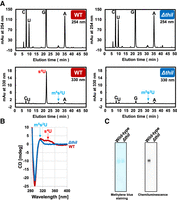
(A) Nucleoside analysis of tRNA fractions from T. thermophilus wild-type (WT) and ΔthiI strains. Absorbances at 254 nm (upper) and 330 nm (lower) were monitored. Transfer RNA fraction from the wild-type strain contains s4U and m5s2U. In contrast, the tRNA fraction from the ΔthiI strain contains only m5s2U. The peak areas of s4U and m5s2U in the lower panels do not represent the molecular ratio between s4U and m5s2U, because the absorption of UV at 330 nm by m5s2U is weak. (B) CD-spectra of tRNA fractions from T. thermophilus wild-type (WT) and ΔthiI strains. The presence of m5s2U in the tRNA fraction from the ΔthiI strain was confirmed. (C) Transfer RNA fractions from T. thermophilus wild-type and ΔthiI strains reacted with MTSEA biotin-XX were separated by 10% PAGE (7 M urea) (left) and visualized by methylene blue staining. The RNAs were blotted onto a nylon membrane and then chemiluminescence derived from bound streptavidin-HRP was monitored.
The MTSEA biotin-XX s4U detection system can be used for analysis of in vitro enzymatic s4U synthesis
Incorporation of a 35S-atom from 35S-labeled l-cysteine into a tRNA transcript is often used in enzymatic analysis of in vitro s4U synthesis (Lauhon 2002; Lauhon et al. 2004). This method is direct, sensitive, and quantitative. However, use of radioisotope-labeled compounds requires a specific facility, and the half-life of 35S is 87.5 d. We tested whether the MTSEA biotin-XX s4U detection system can be used for enzymatic analysis of s4U synthesis. For this purpose, E. coli ThiI and IscS were individually expressed in E. coli cells and purified (Fig. 5A). We prepared E. coli tRNAPhe transcript (Fig. 5B) as a substrate tRNA because this tRNA and its truncated RNAs were previously used in enzymatic analyses of ThiI (Kambampati and Lauhon 2000; Lauhon et al. 2004; Neumann et al. 2014). Because E. coli ThiI and IscS do not possess an Fe-S cluster, the sulfur-transfer reaction is able to be measured under aerobic conditions. When ThiI, IscS, l-cysteine and tRNAPhe transcripts were incubated in the absence of ATP, s4U formation did not occur (Fig. 5C, left). In contrast, when 2 mM ATP was added into the reaction mixture, the MTSEA biotin-XX system detected the formation of s4U in the tRNAPhe transcript (Fig. 5C right). Thus, the MTSEA biotin-XX system can be used for the analysis of in vitro s4U synthesis instead of 35S-labeled cysteine.
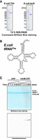
(A) Purified ThiI (2.0 µg; left) and IscS (2.0 µg; right) were analyzed by 15% SDS-PAGE. The gels were stained with Coomassie brilliant blue. (B) Sequence of E. coli tRNAPhe transcript. (C) Formation of s4U in E. coli tRNAPhe transcript by ThiI was detected using the MTSEA biotin-XX s4U detection system. In the absence of ATP, s4U formation did not occur (negative control). In contrast, in the presence of 2 mM ATP, time-dependent s4U formation was observed by the MTSEA biotin-XX s4U detection system.
The MTSEA biotin-XX s4U detection system can visualize newly transcribed tRNAs in eukaryotic cells
MTSEA biotin-XX does not react with sulfur-containing modified nucleosides with the exception of s4U. This finding prompted us to apply the MTSEA biotin-XX s4U detection system for visualization of newly transcribed RNAs. We selected Saccharomyces cerevisiae as a model organism because the sequences of almost all tRNAs from S. cerevisiae have been investigated and s4U is not present in S. cerevisiae tRNAs (Sajek et al. 2020). Figure 6A shows the outline of this experiment. Briefly, S. cerevisiae cells were cultured and pulse-labeled with 4-thiouracil when the optical density at 600 nm reached 0.9. After 15 min, the cells were collected, and total RNA was prepared. Total RNAs were reacted with MTSEA biotin-XX, separated by 10% PAGE (7 M urea) and then electroblotted onto a nylon membrane filter. Chemiluminescence from the filter was monitored using streptavidin-HRP. Figure 6B shows the result of this experiment. In the negative control, which was added water instead of 4-thiouracil, weak chemiluminescence was observed at the large RNA region (left lane in Fig. 6B). This RNA is mainly rRNAs. Although the total RNA was treated with proteinase K to remove proteins, denatured ribosomal proteins probably remained. Notably, chemiluminescence was not detected from the small RNAs (mainly tRNAs) in the negative control sample. Fifteen minutes after the addition of 4-thiouracil, chemiluminescence was clearly detected from tRNAs (right lane in Fig. 6B). Thus, the MTSEA biotin-XX s4U detection system can be used to monitor newly transcribed tRNAs.
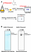
(A) Schematic drawing of detection of newly transcribed RNAs in S. cerevisiae cells by the MTSEA biotin-XX detection system. (B) Total RNAs from the samples without addition of s4U (left lane)- and with addition of s4U (right lane) were separated by 10% PAGE (7 M urea). The gel was stained with methylene blue. 4-Thiouridine in total RNAs was detected by the MTSEA biotin-XX system.
Conclusions
Transfer RNA modification and RNA technology fields require a selective and sensitive s4U detection system. Our experimental results show that the MTSEA biotin-XX s4U detection system is sensitive and quantitative. This system can be used without denaturation; however, addition of a denaturation step improves the limit of detection. The most useful advantage of the MTSEA biotin-XX s4U detection system is that MTSEA biotin-XX reacts only with s4U and not with other sulfur-containing modified nucleosides such as s2U derivatives in tRNAs. This feature is useful for the studies of in vivo tRNA transcription and tRNA modification. Furthermore, the MTSEA biotin-XX s4U detection system can be used to analyze multiple samples in a short time. Moreover, the MTSEA biotin-XX s4U detection system can be used for the enzymatic analysis of s4U formation in tRNA. Therefore, when a radioisotope facility is not available, this system will be useful. Finally, we were able to visualize newly transcribed tRNAs in S. cerevisiae cells using the MTSEA biotin-XX system. Therefore, because MTSEA biotin-XX does not react with sulfur-containing modified nucleosides with the exception of s4U, this system is applicable in the analysis of newly transcribed tRNAs in eukaryotic cells.
MATERIALS AND METHODS
Materials
MTSEA biotin-XX was purchased from Fuji Film-Wako. Hybond-N+ nylon membrane filter was bought from Cytiva. 4-Thiouracil, 4-thiouridine, and s4UTP were obtained from Tokyo Kasei, Sigma, and Jena Bioscience, respectively. Streptavidin-HRP (HRP-conjugated streptavidin) was purchased from Proteintech. Other chemical reagents were of analytical grade.
Preparation of Linear-s4U, Linear-U, and Stem-s4U RNAs
Linear-s4U, Liner-U, and Stem-s4U RNAs were transcribed by T7 RNA polymerase using the same method for preparation of tRNA transcript (Hori 2010). The template DNAs were constructed using the following primers: Linear-s4U F, 5′-GCGAAATTAATACGACTCACTATAGGGACGCGTGCGCAAAG-3′; Linear-s4U R, 5′-CTGCGCTGTCCTGGTCTGCCTTTGCGCACGCGTCCCTATAG-3′; Stem-s4U F, 5′-GCGAAATTAATACGACTCACTATAGGGACGCGTGCGCACGG-3′; Stem-s4U R, 5′-TGCTTCGCGTGCGCTGCCTCCGTGCGCACGCGTCCCTATA-3′. In the case of Linear-s4U and Stem-s4U transcriptions, s4UTP was used instead of UTP. The transcribed RNAs were purified by 10% PAGE (7 M urea).
Determination of reaction time of MTSEA biotin-XX and s4U
Linear-s4U RNA (final concentration, 10 µM) was dissolved in 100 µL of reaction mixture (10 mM HEPES-KOH [pH 7.6], 1 mM EDTA, 20% dimethyl sulfoxide). MTSEA biotin-XX was added into the reaction mixture (final concentration, 82.5 µM). The absorption at 330 nm derived from s4U was measured using an Ultrospec 6300 pro UV/visible spectrophotometer (Amersham Biosciences).
Detection of s4U by chemiluminescence
Linear-s4U and -U RNAs were dissolved in water, denatured by rapid cooling from 80°C to 4°C, and sequentially diluted. The MTSEA biotin-XX was reacted with the diluted RNAs in 250 µL of reaction mixture (10 mM HEPES-KOH [pH 7.6], 1 mM EDTA, 20% dimethyl sulfoxide) at 25°C for 30 min. The RNAs were recovered by phenol/chloroform treatment and ethanol precipitation, dissolved in water, spotted onto a Hybond-N+ nylon membrane filter and fixed by UV at 254 nm irradiation. The RNAs were visualized by methylene blue staining and then the filter was washed with water. The filter was incubated in 10 mL Blocking One solution (code 0395-95, Nacalai Tesque) at room temperature overnight and then washed with 10 mL of TBST buffer (20 mM Tris-HCl [pH 7.6], 140 mM NaCl, 0.1% Tween 20) twice. The filter was dipped into 15 mL of mixture of 95% TBST buffer and 5% Blocking One. An amount of 3 µL of streptavidin-HRP was directly added to the buffer and then incubated at room temperature for 90 min. The filter was washed with 15 mL TBST buffer twice and then washed with 15 mL water. The bound HRP was detected by chemiluminescence using EzWestLumi plus (code WSE-7120S, Atto) and Lumino Graph II EM (code WSE-6270, Atto) according to the manufacture's manual. For Stem-s4U RNA, the RNA (final concentration, 9.2 µM) was annealed in 100 µL annealing buffer (10 mM HEPES-KOH [pH 7.6], 5 mM MgCl2, 100 mM NaCl) by cooling from 80°C to room temperature for 15 min. Denaturation of annealed Stem-s4U RNA was performed by rapid cooling from 80°C to 4°C in the presence of 10 mM EDTA.
Culture of Thermus thermophilus strains
The culture source of Thermus thermophilus wild-type strain (Oshima and Imahori 1974) was a gift from Dr. Tairo Oshima (Kyowa Kako). Construction of the T. thermophilus ΔthiI strain has been previously reported (Shigi et al. 2006). Both strains were cultured in nutrient-rich medium (Tomikawa et al. 2010) at 65°C. The cells were collected when the optical density at 600 nm was reached 0.9, frozen in liquid-nitrogen, and stored at −80°C before use.
Preparation of tRNA fractions from T. thermophilus cells
Total RNA was extracted from the cultured cells using the acid guanidium thiocyanate-phenol–chloroform extraction method (Chomezynski and Sacchi 1987). Transfer RNA fractions were prepared from the total RNA by 10% PAGE (7 M urea).
Nucleoside analysis of tRNA fractions
Modified nucleosides in tRNA fractions were analyzed by the method reported previously (Tomikawa et al. 2010). Briefly, 0.30 A260units of tRNA fraction was digested with 0.5 units Nuclease P1 (Fuji Film-Wako), 20 µg RNase A (Invitrogen) and 0.125 units bacterial alkaline phosphatase (Takara) at 37°C over-night. Nucleosides were analyzed on a Hitachi L-2000 HPLC system (Hitachi) equipped with a Nucleosil 7C18 column (Chemco Plus).
Measurement of CD spectra
Transfer RNA fractions (final concentration 124.2 A260 units/ml) were dissolved in 220 µL of CD buffer (50 mM Tris-HCl [pH 7.6], 5 mM MgCl2, 50 mM KCl). CD spectra were measured on a JASCO J-820 spectropolarimeter equipped with a JASCO PTC-423L thermo-controller at 20°C. Cuvettes with a 1 mm path length were used. The spectra were recorded from 400 nm to 280 nm. The scan speed was 160 nm/min. The spectra shown in this report are the average of five scans.
Detection of s4U in T. thermophilus tRNA fraction
0.20 A260 units of tRNA fractions of T. thermophilus wild-type and ΔthiI strains were reacted with MTSEA biotin-XX, recovered by phenol–chloroform extraction and ethanol precipitation and then separated by 10% PAGE (7 M urea). The gel was stained with methylene blue. The equal amounts of tRNA fractions reacted with MTSEA biotin-XX were blotted onto a Hybond-N+ nylon membrane filter. The filter was incubated in 10 mL Blocking One solution at room temperature overnight and then washed with 10 mL of TBST buffer twice. The filter was dipped into 15 mL of mixture of 95% TBST buffer and 5% Blocking One. A total of 3 µL of streptavidin-HRP was directly added into the buffer and then incubated at room temperature for 90 min. The filter was washed with 15 mL TBST buffer twice and then washed with 15 mL water. The bound HRP was detected by chemiluminescence using EzWestLumi plus and Lumino Graph II EM according to the manufacturer's manual.
Construction of E. coli ThiI and IscS expression systems
Escherichia coli thiI and iscS genes were amplified by polymerase chain reaction (PCR) from the genomes of E. coli DH5α (Toyobo) and BL21 (DE3) Rosetta 2 (Novagen) strains, respectively, using the following primers: Eco thiI F, 5′-GAAGGAGATATACATATGAAGTTTATCATTAAATTGTTCCCG-3′; Eco thiI R, 5′-GAGCTCGAATTCGGATCCTCATTACGGGCGATATACCTTCACATT-3′; Eco iscS F, 5′-GAAGGAGATATACATATGAAATTACCGATTTATCTCGACTAC-3′; Eco iscS R, 5′-GAGCTCGAATTCGGATCCTCATTAATGATGAGCCCATTCGATGCT-3′. The amplified DNAs were purified using 1% agarose gel electrophoresis and then inserted between NdeI and BamHI sites of pET30a plasmid vector (Novagene) using NEBuilder HiFi DNA assembly Master Mix (New England Biolab).
Expressions of E. coli ThiI and IscS
The constructed plasmids (pET-30a-EcothiI and pET-30a EcoiscS) were individually introduced into E. coli BL21 (DE3) Rosetta 2 strain. The cells were cultured in 1 L of LB medium at 37°C. When the optical density at 600 nm reached 0.6, isopropyl β-D-thiogalactopyranoside was added into the medium (final concentration, 1 mM) and then cultivation was continued for 4 h. The cells were collected by centrifugation at 4320g at 4°C for 20 min, frozen in liquid-nitrogen and stored at −80°C before use.
Purification of E. coli ThiI
Wet cells (0.9 g) were suspended in 19 mL of buffer A (50 mM Tris-HCl [pH 7.6], 5 mM MgCl2, 50 mM KCl, 6 mM 2-mercaptethanol, 5% glycerol) supplemented with Protease Inhibitor Cocktail (code: 03989-34, Nacalai Tesque), and then disrupted with an ultrasonic disruptor (model VCX-500, Sonics and Materials. Inc). The supernatant was collected by centrifugation at 38,900g at 4°C for 20 min, and then loaded onto a Q-Sepharose Fast Flow column (column volume 5 mL, Cytiva). ThiI was eluted by a linear gradient from 50 mM to 1200 mM KCl in buffer A. The ThiI fractions were assessed by 15% SDS-PAGE and combined. The KCl concentration in the sample was adjusted to around 40 mM by dilution with buffer A without KCl. The sample was loaded onto a HiTrap Heparin HP column (column volume 5 mL, Cytiva). ThiI was eluted by a linear gradient from 50 mM to 1200 mM KCl in buffer A. The ThiI fractions were assessed by 15% SDS-PAGE, combined, and concentrated using a Vivaspin Turbo 15 filter device (Sartorius). The concentrated sample was loaded onto a Superdex-200 prep grade gel-filtration column (column volume 120 mL, Cytiva), which was equilibrated in buffer B (50 mM Tris-HCl [pH 7.6], 5 mM MgCl2, 400 mM KCl, 5 mM dithiothreitol). The ThiI fractions were combined, and then loaded onto a Bio-Gel Hydroxyapatite HP column (Bio-Rad), which was equilibrated in buffer A. ThiI was eluted in 25 mL of buffer C (50 mM potassium phosphate buffer [pH 8.0], 1 M KCl, 5 mM MgCl2, 6 mM 2-mercaptoethanol). The ThiI fractions were combined and then the buffer was exchanged to buffer A in a Vivaspin Turbo 15 filter device. Glycerol was added (final concentration, 50%). The purified ThiI was stored at −30°C.
Purification of E. coli IscS
Wet cells (0.7 g) were suspended in 19 mL of buffer A supplemented with Protease Inhibitor Cocktail, and then disrupted with an ultrasonic disruptor. The supernatant was collected by centrifugation at 38,900g at 4°C for 20 min, and then loaded onto a Q-Sepharose Fast Flow column. IscS was eluted by a linear gradient from 50 mM to 1200 mM KCl in buffer A. The IscS fractions were assessed by 15% SDS-PAGE and combined. The KCl concentration in the sample was adjusted to ∼40 mM by dilution with buffer A without KCl. The sample was loaded onto a HiTrap Heparin HP column. IscS was eluted by a linear gradient from 50 mM to 1200 mM KCl in buffer A. The IscS fractions were assessed by 15% SDS-PAGE, combined, and concentrated using a Vivaspin Turbo 15 filter device. The concentrated sample was loaded onto a Superdex-75 prep grade gel-filtration column (column volume 120 mL, Cytiva), which was equilibrated in buffer A containing 200 mM KCl. The eluted IscS fractions were combined and concentrated using a Vivaspin Turbo 15 filter device. Glycerol was added (final concentration, 50%). The purified IscS was stored at −30°C.
Preparation of E. coli tRNAPhe transcript
Escherichia coli tRNAPhe transcript was prepared by T7 RNA polymerase as described in the reference (Hori 2010). The transcript was purified by 10% PAGE (7 M urea).
Reaction of ThiI, IscS, E. coli tRNAPhe transcript, and l-cysteine in the presence and absence of ATP
An amount of 1.0 µM ThiI, 1.0 µM IscS, 0.1 A260units E. coli tRNAPhe transcript, and 1 mM l-cysteine were mixed in 50 µL of ThiI-IscS buffer (50 mM Tris-HCl [pH 7.6], 5 mM MgCl2, 50 mM KCl, 100 µM 2-mercaptoethanol, 1 mM pyridoxal phosphate) in the presence and absence of 2 mM ATP. The mixtures were incubated at 37°C. The reaction was stopped by addition of 50 µL phenol–chloroform at 0, 1, 2, 5, 10, and 15 min. The RNAs were recovered by ethanol precipitation and then s4U was detected by the same method as described in the Detection of s4U in T. thermophilus tRNA fraction section.
Saccharomyces cerevisiae strain
Saccharomyces cerevisiae BY4743 strain was purchased from National BioResource Project - Yeast (National Institute of Genetics).
Pulse-labeling of S. cerevisiae RNAs by 4-thiouracil
Saccharomyces cerevisiae cells were cultured at 30°C in 200 mL YPD medium (4 g glucose, 4 g peptone, 2 g yeast extract per 200 mL). When the optical density at 600 nm reached 0.9, 4-thiouracil (final concentration 5 mM) was added. Negative control was prepared by addition of water instead of 4-thiouracil. After 15 min, the cells were collected by centrifugation at 4320g at 4°C for 20 min. Total RNAs were prepared by the acid guanidium thiocyanate-phenol–chloroform extraction method (Chomezynski and Sacchi 1987). Total RNA (1.50 A260 units each) was further treated with 5 µg proteinase K in 100 µL of reaction mixture (10 mM Tris-HCl [pH 7.6], 10 mM EDTA, 0.5% SDS) at 50°C for 1 h. Total RNAs were extracted with phenol–chloroform, recovered by ethanol precipitation, and then reacted with MTSEA biotin-XX. The total RNA (0.60 A260 units each) was separated by 10% PAGE (7 M urea) and then blotted onto a Hybond-N+ nylon membrane filter. Chemiluminescence was detected by the same method as described in the detection of s4U in T. thermophilus tRNA fraction section. Escherichia coli tRNAPhe transcript (0.15 A260units), which was modified by 1.0 µM ThiI and 1.0 µM IscS in the presence of 1 mM l-cysteine and 2 mM ATP at 37°C for 2 h, was used as a marker.
SUPPLEMENTAL MATERIAL
Supplemental material is available for this article.
ACKNOWLEDGMENTS
This work was supported by a Grant-in-Aid for Scientific Research from the Japan Society for the Promotion of Science (JSPS) (20H03211 to H.H. and 21H02436 to N.S.).
Footnotes
-
Article is online at http://www.rnajournal.org/cgi/doi/10.1261/rna.079445.122.
- Received September 7, 2022.
- Accepted November 14, 2022.
This article is distributed exclusively by the RNA Society for the first 12 months after the full-issue publication date (see http://rnajournal.cshlp.org/site/misc/terms.xhtml). After 12 months, it is available under a Creative Commons License (Attribution-NonCommercial 4.0 International), as described at http://creativecommons.org/licenses/by-nc/4.0/.




