


Cover image
On the cover
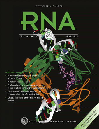
Cover Illustration: Crystal structure of a human IgG–aptamer complex (Protein Data Bank code: 3agv; Nomura Y, Sugiyama S, Sakamoto T, Miyakawa S, Adachi H, Takano K, Murakami S, Inoue T, Mori Y, Nakamura Y, et al. 2010. Conformational plasticity of RNA for target recognition as revealed by the 2.15 Å crystal structure of a human IgG–aptamer complex. Nucleic Acids Res 38: 7822–7829). Image details show protein—ribbon representation, chain-specific coloring; aptamer—ribbon-plate representation, nucleotide-specific coloring, A—red, G—green, modified nucleotides—cyan; carbohydrates—stick representation, violet; Ca ions—ball representation, blue. The image was generated with the Accelrys Discovery Studio Visualizer. Cover image provided by the Jena Library of Biological Macromolecules—JenaLib (www.fli-leibniz.de/IMAGE.html).


Molecular Cloning
The New Edition
Buy Now and Save 30%
Paperback only
(limited time offer)

|
Molecular Cloning: A Laboratory Manual (Fourth Edition) |