


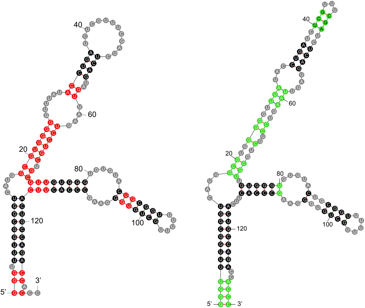
FIGURE 1.
Two secondary structures for the 5S rRNA from the Archaeon, Methanothermobacter thermautotrophicus (strain δ-H; variant 2). (Left) The accepted phylogenetic structure (Szymanski et al. 2002). (Right) A minimum free energy (mfe) structure computed by UNAFold (Markham and Zuker 2008). Each base is drawn within a colored circle: (black circles) base pairs that are common to both structures, (red circles) base pairs in the phylogenetic structure that do not occur in the mfe structure, (green circles) base pairs in the mfe structure that are not in the phylogenetic structure, (gray circles) single-stranded bases.




