


Plasticity of the RNA kink turn structural motif
Abstract
The kink turn (K-turn) is an RNA structural motif found in many biologically significant RNAs. While most examples of the K-turn have a similar fold, the crystal structure of the Azoarcus group I intron revealed a novel RNA conformation, a reverse kink turn bent in the direction opposite that of a consensus K-turn. The reverse K-turn is bent toward the major grooves rather than the minor grooves of the flanking helices, yet the sequence differs from the K-turn consensus by only a single nucleotide. Here we demonstrate that the reverse bend direction is not solely defined by internal sequence elements, but is instead affected by structural elements external to the K-turn. It bends toward the major groove under the direction of a tetraloop–tetraloop receptor. The ability of one sequence to form two distinct structures demonstrates the inherent plasticity of the K-turn sequence. Such plasticity suggests that the K-turn is not a primary element in RNA folding, but instead is shaped by other structural elements within the RNA or ribonucleoprotein assembly.
Keywords
INTRODUCTION
Analysis of structural elements that provide stability to global RNA complexes is important for understanding the rules of RNA folding. The kink turn (K-turn) is a well-studied RNA motif that forms both structured and flexible conformations within larger RNA molecules (Matsumura et al. 2003). The plasticity of the K-turn motif and the ability to analyze its properties at an atomic level make it a good model for studying RNA structure (Turner et al. 2005).
The K-turn was first described in the large ribosomal subunit of Haloarcula marismortui (Klein et al. 2001). Six examples of this motif are present within the H. marismortui 50S subunit, and two are present in the Thermus thermophilus 30S subunit (Ban et al. 2000; Schluenzen et al. 2000; Wimberly et al. 2000). Other K-turns are located in non-ribosomal structures such as riboswitches, small nucleolar RNAs, and ribozymes (Nottrott et al. 1999; Adams et al. 2004; Montange and Batey 2006; Blouin and Lafontaine 2007). The K-turn is composed of 15 nucleotides (nt) that form a helix–internal-loop–helix structure in duplex RNA (Fig. 1). Ten of these 15 nt make up the K-turn's consensus secondary structure (Klein et al. 2001). A standard K-turn has a canonical helix (C-stem), formed from C–G pairs, followed by a 3-nt, asymmetric internal loop that ends with a noncanonical helix (NC-stem) of G·A mispairs. Tertiary interactions to the two closing G·A mispairs facilitate the K-turn bend and include a stabilizing A-minor motif contact to the A in the first G·A pair. The three nucleotides within the asymmetric loop create the kink in the phosphodiester backbone of the RNA. Nearly every K-turn adheres to the consensus secondary structure and forms a 120° bend in the helix toward the minor groove (Klein et al. 2001).
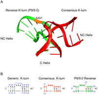
(A) Reverse K-turn bent toward the major grooves (green) and consensus K-turn bent toward the minor grooves (red) with position A201 of the Azoarcus intron highlighted in orange. (B) Generic K-turn numbering system (black) as compared with consensus (red) and reverse K-turns (green) with position A201 highlighted in orange.
Unlike other K-turns, the crystal structure of the Azoarcus group I intron revealed a helix–loop–helix element that has many secondary structural features of a consensus K-turn, yet it is bent in the opposite direction (Adams et al. 2004). Similar to the consensus K-turn, the reverse K-turn has a 3-nt asymmetric bulge and is flanked on both sides by C- and NC-stems. A 90° bend forms at the asymmetric loop between the P9.0 and P9. The only difference in sequence between the K-turn consensus and that of the reverse K-turn is a G-to-A change (nucleotide 201, Azoarcus numbering) in the first base pair of the NC-stem, resulting in an A·A instead of a G·A mispair. In a standard K-turn, the 5′-most nucleotide of the loop stacks on the C-stem, allowing the second nucleotide in the bulge to stack on the NC-stem, and the third nucleotide to be displaced into solution. In the reverse K-turn, all three residues stack beneath either the C- or NC-stem with no residue extended into solution. The absence of the A-minor interaction normally present in consensus K-turns suggests that the tertiary elements that stabilize the reverse K-turn differ from those that stabilize a standard K-turn. In the reverse K-turn a hydrogen bond is observed between the Hoogsteen edge of the middle unpaired residue (C199) and a nonbridging phosphate oxygen of the NC-stem backbone. A Mg2+ ion may aid in the alignment of the NC-stem phosphate backbone against the C-stem major groove (Strobel et al. 2004).
Given that two similar sequences bend in opposite directions, it raises these questions: Is the bend direction of the reverse K-turn dictated by sequence, tertiary structure interactions, or a combination of both? Is the single change from a G to an A at position 201 responsible for the change in bend direction? Or does it result from a specific sequence combination within the asymmetric bulge? Studies of K-turns, such as the well-studied ribosomal Kt-7, indicate that the two conserved G·A mispairs of the NC helix and the hydrogen-bonding interactions they make are critical for the correct folding and stabilization of a standard K-turn (Liu and Lilley 2007; Turner and Lilley 2008). It may be possible that the reverse K-turn has different internal sequence requirements that specify its alternate conformation.
If sequence is insufficient to specify K-turn direction, what other factors and tertiary interactions are responsible for directing the bend of the K-turn toward the major grooves? Many K-turns serve as protein recognition elements (Klein et al. 2001; Brodersen et al. 2002). Binding of proteins, such as the 15.5K protein to box C/D snoRNA, can influence whether a K-turn remains a flexible loop or becomes locked into a more rigid conformation (Cojocaru et al. 2005; Szewczak et al. 2005). Within the group I intron, a tetraloop–tetraloop receptor (TL-TLR) interaction has been shown to be energetically favorable for stabilization and folding (Rangan et al. 2003). Could this tertiary element play a defining role in K-turn bend direction? In this scenario, the reverse K-turn would serve only as a flexible hinge that is directed toward the major groove by the TL-TLR interaction.
Using biochemical techniques and crystallographic analysis, we determined if mutation of the wild-type reverse K-turn sequence to a consensus K-turn sequence produces a change in bend direction. We further examined the role of both internal and external sequence elements to specify the bend direction of the Azoarcus group I intron K-turn. The data suggest that the K-turn is a malleable element of RNA structure such that two distinct subclasses of tertiary structure can be derived from a single primary nucleic acid sequence.
RESULTS
We explored the sequence requirements of the reverse kink turn using both kinetic and structural analysis. All studies were performed on the asymmetric loop in the P9.0 helix within the Azoarcus group I intron. We measured splicing rates using the ribozyme form of the Azoarcus intron that catalyzes the attack of the 3′-hydroxyl of exogenous GMP on the oligonucleotide substrate CAUAAA (Fig. 2A). A construct in which P9.0 is fully base-paired (eliminating the asymmetric loop and noncanonical pairings) was developed to ascertain the effect of complete disruption of the reverse K-turn and consequent tertiary interactions such as the docking of the TL in its receptor (Fig. 2C). However, under standard reaction conditions, there was less than a fourfold rate difference between wild-type (WT) and the fully base-paired RNA (BP). This small effect was anticipated because P9.0 is a peripheral element and not explicitly required for splicing activity. In order to highlight the role of P9.0 folding in splicing, we included 1 M urea in the reaction condition, which resulted in greater than a 100-fold rate difference between the WT and the BP ribozymes (Fig. 3). This amount of denaturant has a marginal effect on the wild-type ribozyme, which remains competent for splicing in 5 M urea and retains some activity even in 7.5 M urea (Tanner and Cech 1996). The inclusion of 1 M urea in the reaction buffer provided a simple system to assay the effects of P9.0 mutations on bend direction and flexibility.
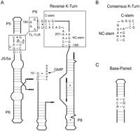
(A) Schematic representation of the Azoarcus group I intron used in kinetic assays. The reverse K-turn sequence (boxed region) differs from a consensus K-turn at position A201. A TL-TLR interaction forms between P9 and J5/5a. Exogenous GMP is added in trans, and cleavage of the 5′ exon (represented by CAUAAA) is used to measure splicing activity. (B) Consensus K-turn sequence with G201. (C) Secondary structure of the base-paired construct.
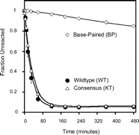
Reaction rates for wild-type (WT), consensus (KT), and base-paired (BP) Azoarcus constructs. The fraction of uncleaved substrate is plotted as a function of time with final rates determined by fitting the curves to a first-order rate equation.
Mutation to the consensus K-turn sequence
The only difference between the reverse K-turn sequence and the consensus K-turn sequence is a single nucleotide in the NC-stem; an A in the reverse K-turn is a G in the canonical sequence. We compared the activity of the WT ribozyme with one in which the sequence was mutated to that of a consensus K-turn (KT ribozyme with an A201G mutation) under the slightly destabilizing reaction conditions described above. The WT ribozyme reacted with a rate of 39 × 10−3 min−1, while the KT ribozyme was negligibly slower, reacting at a rate of 33 × 10−3 min−1 (Table 1). This suggests that the intron remains properly folded with P9.0 bent toward the major groove despite containing a consensus K-turn sequence that would be predicted to bend in the opposite direction.
First-order rate kinetics for Azoarcus group I intron mutants
Mutations within the internal asymmetric loop
The three nucleotides (residues 198–200) in the asymmetric loop of a standard K-turn do not display any sequence conservation and are not expected to be defining elements for the consensus K-turn. To determine if these nucleotides define an obligate reverse K-turn subclass, we mutated the nucleotides in the asymmetric loop. Of particular interest is nucleotide C199, which appears to form a hydrogen bond with the phosphate of G180 in the NC-stem. This interaction could facilitate the close stacking of bases under the C- or NC-helix, helping to stabilize the reverse K-turn bend. Constructs WTC199U and KTC199U disrupt this potential hydrogen bond in the WT and KT backgrounds, respectively. However, the C199U mutation had a small effect on the rate in either context (Table 1). These data suggest that the internal hydrogen-bonding interaction plays at most only a minor role in stabilizing the conformation of the reverse K-turn and is not the defining element for the bend direction.
The asymmetric loop of the reverse K-turn is a relatively A-rich region with adenosines at the first and third nucleotides. Four additional constructs were developed to disrupt the two other positions in the asymmetric loop (constructs WTA198U, (KTA198U, WTA200U, and KTA200U mutate the adenosines at positions 198 and 200 in both the WT and KT backgrounds). For all constructs, the rates are within twofold of the wild-type values. These data further suggest that the three nucleotides in the asymmetric loop, the primary site of the bend, are not a significant determinant of K-turn bend direction. The sequence of the Azoarcus group I intron asymmetric loop does not define a reverse K-turn subclass.
Mutation of external elements
Given that sequence elements within the reverse K-turn do not appear to define bend direction, we next explored the role of external structural factors. We were particularly interested in the possibility that a TL-TLR interaction would have a strong influence on the reverse K-turn bend direction. The tetraloop at the end of P9 interacts with a tetraloop receptor within P5 of this intron (Fig. 2A). We mutated this GAAA tetraloop to UUGG in both the WT and KT backgrounds (WTUUGG and KTUUGG). This resulted in 740- and 925-fold losses in the rate of cleavage for the two constructs. We also examined a more conservative mutation of the tetraloop, A190C, which changes the GAAA tetraloop to GCAA. The result is a stable GNRA tetraloop, but the tetraloop forms a significantly less stable interaction with the receptor (Costa and Michel 1997; Murphy and Cech 1993). We found a moderate fivefold rate effect in the wild-type background (WTA190C), but an almost 30-fold effect for the RNA with mutations in both external and internal regions (KTA190C). The combination of internal and external mutations appears to destabilize the intron more than either point mutation alone.
Structure determination of the consensus sequence
To confirm that we made the proper structural interpretation of this kinetic result, we crystallized the A201G mutant RNA (KT) and obtained the structure by molecular replacement. The construct was assembled in a manner similar to that used to obtain the original Azoarcus group I intron crystal structure (Adams et al. 2004). A 22-residue oligonucleotide, dcircm, which represented the 3′-exon and the 3′-end of the intron and included the A201G point mutation, was crystallized with the 5′-exon CAT and the remainder of the intron produced as an RNA transcript. We obtained a 4.2 Å resolution structure of the mutant group I intron in the same space group (P4122) as previously reported for other Azoarcus intron structures (Table 2; Adams et al. 2004; Stahley and Strobel 2005; Lipchock and Strobel 2008). Both the mutant and the original structures have the same overall architecture (Fig. 4). Although the resolution was limited, there was no observable change in conformation of the phosphate backbone of the P9 helix resulting from the point mutation. Thus, despite a consensus sequence, the P9.0 helix remained bent toward the major grooves. This provides structural support to the kinetic data and suggests that a consensus K-turn sequence can fold in the opposite direction within the context of the Azoarcus ribozyme.
Crystallographic data collection and refinement statistics
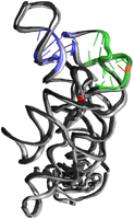
Comparison of a crystal structure of an Azoarcus mutant with a consensus K-turn sequence (green/orange) to the original Azoarcus intron structure (gray). The P9-J5/5a tetraloop–tetraloop receptor interaction is highlighted (blue) as well as a scissile phosphate in the intron core (red).
DISCUSSION
This study demonstrates that the K-turn motif is an example of a plastic secondary structural element that can adopt multiple conformations in the presence of different external factors. We explored the sequence and structural determinants of the reverse K-turn within the context of the Azoarcus ribozyme and found that docking of a TL-TLR can mold a consensus K-turn into an unusual bend direction. The ability of the K-turn to form two distinct, yet stable structures from the same primary sequence supports the proposal that K-turns are not principal organizing elements of RNA structure (Goody et al. 2004; Turner et al. 2005). While other internal loop motifs, such as the bulged-G motif, are defined as rigid regions (Correll et al. 2003), K-turns are dynamic with kinked and unkinked conformations influenced by metal ion concentration, tertiary interactions, and protein binding (Nottrott et al. 1999; Goody et al. 2004).
A tetraloop–tetraloop receptor interaction stabilizes the reverse K-turn bend direction
Solution studies of isolated K-turn motifs show flexible RNAs that are able to form tightly kinked or extended conformations in the presence or absence of metal ions (Goody et al. 2004). Other K-turns, like many in the ribosome, rely on external ligands and tertiary interactions to mold their conformations (Matsumura et al. 2003). Because K-turns can be shaped by external elements we investigated the role of the TL-TLR interaction between P5 and P9 in the bend direction of the reverse K-turn sequence. We observed that the disrupted tetraloop mutants had the most significant impact on reaction rate, which is consistent with the conclusion that mutations to the GAAA tetraloop of L9 disrupted proper intron folding (Rangan et al. 2003). In fact, it was only when this interaction was lost that we were able to observe any effect from mutation of the reverse K-turn to the consensus sequence.
This view of the K-turn is similar to the bent sequence within the P4–P6 domain of the Tetrahymena group I intron, which also requires external tertiary interactions in order to adopt a bend (Cate et al. 1996a,b). The P4–P6 domain consists of 160 nucleotides and forms an independent tertiary structure including a 180° hinge at J5/5a (Szewczak and Cech 1997; Cate et al. 1996a,b). To fold correctly, the motif requires a TL-TLR and A-minor motif interactions. The nucleotide sequence that makes up the J5/5a hinge has only a minor role in specifying folding (Szewczak and Cech 1997). Similar to the J5/5a hinge, we found that the reverse K-turn loop nucleotides do not direct folding of the K-turn in the Azoarcus group I intron; instead, a TL-TLR interaction is the main determinant of the reverse K-turn bending, and the reverse K-turn is simply a flexible region that can be molded into a major groove bend.
In the hierarchy of RNA folding, a TL-TLR has a dominant role in structure stabilization relative to the KT-bend direction. The plasticity of the reverse K-turn enables it to be shaped in different conformations using distal tertiary contacts and protein binding. While it is certainly possible to design RNA to have a K-turn bend, the actual direction of that bend may be more difficult to predict suggesting that the knowledge of structural plasticity for an RNA secondary structure could enhance RNA tertiary structure prediction.
MATERIALS AND METHODS
Assembly of kinetic assay constructs
The WT reverse sequence template consisted of an Azoarcus group I intron construct (L-6 C204) that terminated at nucleotide position C204. Twelve additional constructs used in kinetic experiments were generated using site-directed mutagenesis (QuikChange, Stratagene) with the WT construct serving as template DNA (Table 1).
In vitro transcription
DNA was prepared for in vitro transcription by performing an EarI restriction enzyme digestion. RNA was transcribed using T7 RNA polymerase and purified by electrophoresis in a 6% polyacrylamide gel containing 7 M urea. Following an overnight elution step in water, the RNAs were concentrated to 0.1–1 mg/mL using Amicon Centrifugal Filter devices.
Short oligonucleotide radiolabeling
A small oligonucleotide was 5′-end-radiolabeled in a 10-μL reaction with [γ-32P]ATP using T4 PNK. Reagents were incubated for 1 h at 37°C and quenched by the addition of formamide loading buffer (95% formamide, 2.5 mM EDTA, 0.1% bromophenol blue, and 0.1% xylene cyanol). The labeled oligonucleotide was purified by 20% non-denaturing PAGE, and the labeled oligonucleotide was eluted overnight in water.
Kinetic measurements
Kinetic reactions were performed to measure the rate of splicing activity for each construct. Reactions contained 3 μM RNA,
1× kinetic buffer [3 mM MgCl2, 25 mM MES at pH 6.0, and 1 mM Mn(OAc)2], 5 mM exogenous guanosine monophosphate (GMP), 1 M urea, and trace amounts of 32P-radiolabeled CAUAAA. Reaction mixtures were preincubated for 2 min at 50°C before the addition of labeled oligonucleotide
and exogenous GMP. Time points were taken by removing 2 μL of the reaction mixture and quenching with 8 μL of formamide loading
buffer. A 20% denaturing polyacrylamide gel was used to separate the unligated substrates from the ligated products. RNA fragments
were visualized using a Storm PhosphorImager (GE Healthcare), and data were quantized using ImageQuant (GE Healthcare). For
each construct, a reaction rate was found by plotting the fraction of unligated product versus time and fitting the data with
KaleidaGraph to an exponential decay endpoint using Equation 1: where funreact = fraction unreacted; Funreact = fraction unreactive.
Crystallization construct assembly and structure determination
A single crystallization construct was assembled from an RNA transcript (UP62) and two synthetic oligonucleotides (CAT and dcircm; Dharmacon). The oligonucleotide, dcircm, represents the 3′-end of the intron and the 3′ exon. It contains a G at position 201, instead of A, to create the KT sequence, and a deoxyG at position 206. Oligonucleotides were deprotected according to the manufacturer's protocol. The UP62 RNA (120 μM) and dcircm (150 μM) were combined and heated for 2 min to 50°C in 10 mM sodium cacodylate (pH 6.5) and 15 mM Mg(OAc)2. After slow cooling for 15 min, CAT (150 μM) and the RNA binding domain of the U1A protein (140 μM) were added. Hanging drop crystallization trials were performed as described previously (Adams et al. 2004). After 3 h, streak seeding was performed using crystals containing an unmutated oligonucleotide. The average crystal size was 120 μm × 100 μm × 100 μm, and crystals typically appeared 12 h after seeding. Crystals were stabilized in 30% MPD, 50 mM sodium cacodylate (pH 6.8), 10 mM Mg(OAc)2, 10 mM KOAc, and 0.2 mM Co(NH3)63+; and were flash frozen in liquid nitrogen.
Diffraction data were indexed and scaled using HKL2000, and the Azoarcus deoxy pre-2S group I intron structure (PDB ID 1U6B) was used to perform molecular replacement to generate a model of the mutant RNA (Otwinoski and Minor 1997; Adams et al. 2004). As previously reported, this construct yielded crystals of space group P4122. Modeling and rigid body refinement of the structure was performed with COOT and REFMAC5 (Winn et al. 2001; Emsley and Cowtan 2004).
Protein Data Bank entry
Atomic coordinates and structure factors have been deposited in the Protein Data Bank, http://www.rcsb.org, under PDB ID 3IIN.
ACKNOWLEDGMENTS
We thank the Beamline staff at the National Synchrotron Light Sourc (NSLS) for assistance with X-ray data collection. This work was supported by NIH PO1 grant GM022778 to S.A.S.
Footnotes
-
Reprint requests to: Scott A. Strobel, Department of Molecular Biophysics and Biochemistry, Yale University, New Haven, CT 06520-8114, USA; e-mail: scott.strobel{at}yale.edu; fax: (203) 432-5767.
-
Article published online ahead of print. Article and publication date are at http://www.rnajournal.org/cgi/doi/10.1261/rna.1883810.
-
- Received August 17, 2009.
- Accepted December 9, 2009.
- Copyright © 2010 RNA Society




