


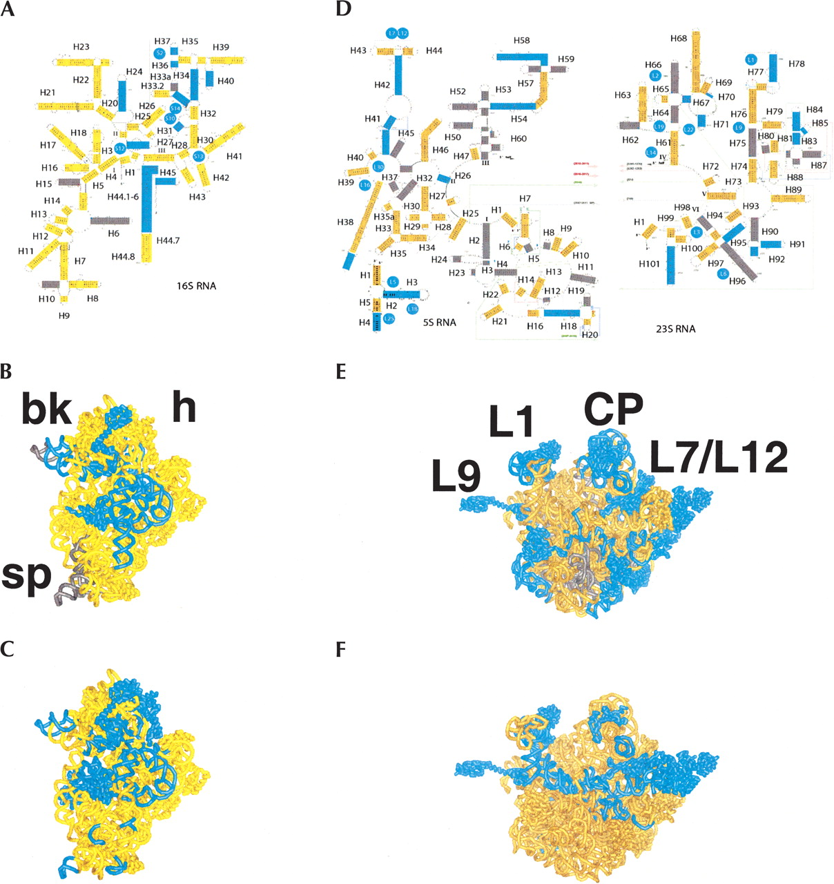
Distribution of regions for which no crosslinking, footprinting, or cleavage information was available (dim gray in A, B, D and E) and ≥25% of the data available was in disagreement with crystal structures (blue in A, B, D and E). (A) Ratios of mismatches mapped into secondary structure of ribosomal 16S RNA. Proteins bearing >25% mismatches in crystallography vs. biochemistry comparisons (see the text) are shown as blue circles placed approximately near binding sites in RNA. (B) Interface of the 30S subunit. The same color scheme as in A, but rendered in three dimensions. (C) Ligand-binding regions (Pioletti et al. 2001; Yusupov et al. 2001; Klaholz et al. 2003; Rawat et al. 2003) and regions found to be flexible in comparative structural studies (see Matadeen et al. 1999; Yusupov et al. 2001; Ogle et al. 2002; Klaholz et al. 2003; Rawat et al. 2003) mapped into crystal structures of the 30S subunit (PDB identification: 1GIX; Yusupov et al. 2001). (D) Secondary structure diagram of the 23S RNA, 5S RNA, and associated proteins, colored in dark gold if data available for the region generally were in good agreement (<25% of mismatch); blue, >25% of mismatches; and gray, no data in our data set. (E) Interface of the 50S subunit. The same color scheme as in D, but rendered in three dimensions. (F) Ligand-binding regions and regions found to be flexible in comparative structural studies (Matadeen et al. 1999; Yusupov et al. 2001; Klaholz et al. 2003; Rawat et al. 2003) mapped into crystal structures of the 50S subunit (PDB identification: 1GIY; Yusupov et al. 2001). Landmarks show macroscopic features of the 30S and 50S subunits known from electron microscopy: h indicates head; bk, beak; sp, spur; and CP, central protuberance (where the 5S RNA is located); as well as proteins L1, L9, and L7/L12 dimer.




