


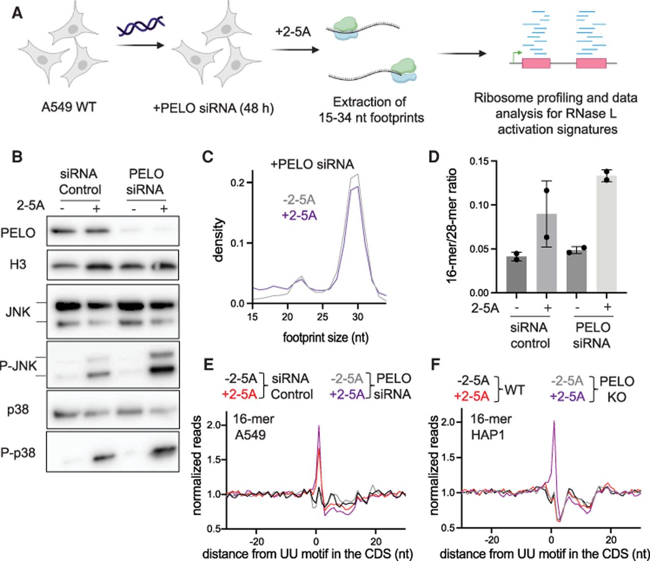
PELO rescues stalled ribosomes at the 3′ end of mRNA fragments during RNase L activation. (A) Schematic representation of experiments. A549 WT cells were treated with PELO siRNA or control siRNA for 48 h followed by transfection with 2-5A or lipofectamine alone (−2-5A). Cells were collected, and ribosome profiling for 15–34 nt footprints was carried out. (B) Western blot assay levels of protein during RNase L activation and PELO knockdown. Comparison of PELO abundance (upper panels) shows effectiveness of knockdown. JNK and p38 phosphorylation (increased by 2-5A) were followed by western blotting with their respective phospho-antibody while also monitoring for total protein levels (lower panels). (C) Normalized length distribution of transcriptome-mapped ribosome profiling reads (15–34 nt): 12,216,111 reads for −2-5A and 3,174,541 reads for +2-5A. (D) To analyze the effects of RNase L activation (2-5A treatment), the ratio of 16-mers (15–18 nt) to 28-mers (25–34 nt) was computed and found to be more significant when PELO was knocked down (P = 0.002 for KD and 0.11 for control comparisons by t-test). Whiskers represent standard deviation between replicates. (E,F) Position average plot of 3′ ends of 16-mers near RNase L cleavage motif (UU) in PELO KD A549 cells (E) and PELO KO HAP1 cells (F). Control data in D and E same as in Figure 4.




