


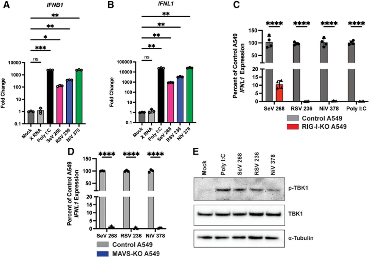
cbVG from RSV and NiV induce MAVS-dependent IFN expression. (A,B) Quantitative PCR (qPCR) for (A) type I interferon (IFNB1) and (B) type III interferon (IFNL1) expression by A549 cells at 6 h post transfection (hpt) with 5 pmol of HCV X RNA, SeV cbVG 268, RSV cbVG 236, or NiV cbVG 378 or 100 ng of poly I:C. One-way ANOVA with Tukey's HSD post hoc test was performed for statistical analysis. (ns) P > 0.05, (*) P < 0.05, (**) P < 0.01, (***) P < 0.001. Data are represented as fold change over Mock. (C) qPCR for IFNL1 of control or RIG-I-KO A549 cells at 6 hpt with 5 pmol of SeV cbVG 268, RSV cbVG 236, or NiV cbVG 378 or 100 ng of poly I:C. One-way ANOVA with Tukey's HSD post hoc test was performed for statistical analysis. (****) P < 0.0001. (D) qPCR for IFNL1 of control or MAVS-KO A549 cells at 6 hpt with 5 pmol of SeV cbVG 268, RSV cbVG 236, or NiV cbVG 378. One-way ANOVA with Tukey's HSD post hoc test was performed for statistical analysis. (****) P < 0.0001. Data reported are biological replicates. (E) Western blots of cell lysates collected from A549 cells 6 hpt with 5 pmol of SeV cbVG 268, RSV cbVG 236, or NiV cbVG 378 or 100 ng of poly I:C. Blots were stained for phosphorylated-TBK1, TBK1, and α-tubulin.




