


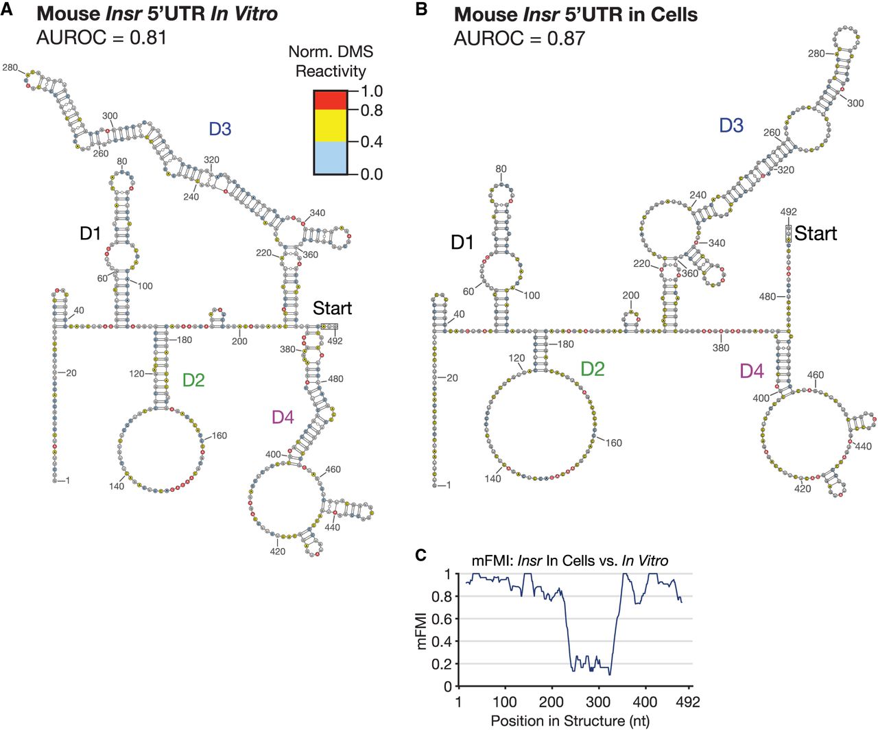
FIGURE 4.
Modeling RNA secondary structure of the mouse Insr 5′UTR with DMS-MaPseq. (A) Model of the mouse Insr 5′UTR constrained by DMS-MaPseq in vitro (AUROC = 0.81), probed in tandem with the viral IRESs in Figures 2 and 3 and modeled using the same prediction parameters (see Materials and Methods). Exact domain divisions are depicted using this model in Supplemental Figure S3A. (B) Model constrained by DMS probing in live cells (AUROC = 0.87). (C) mFMI between the two predicted structures in A and B.




