


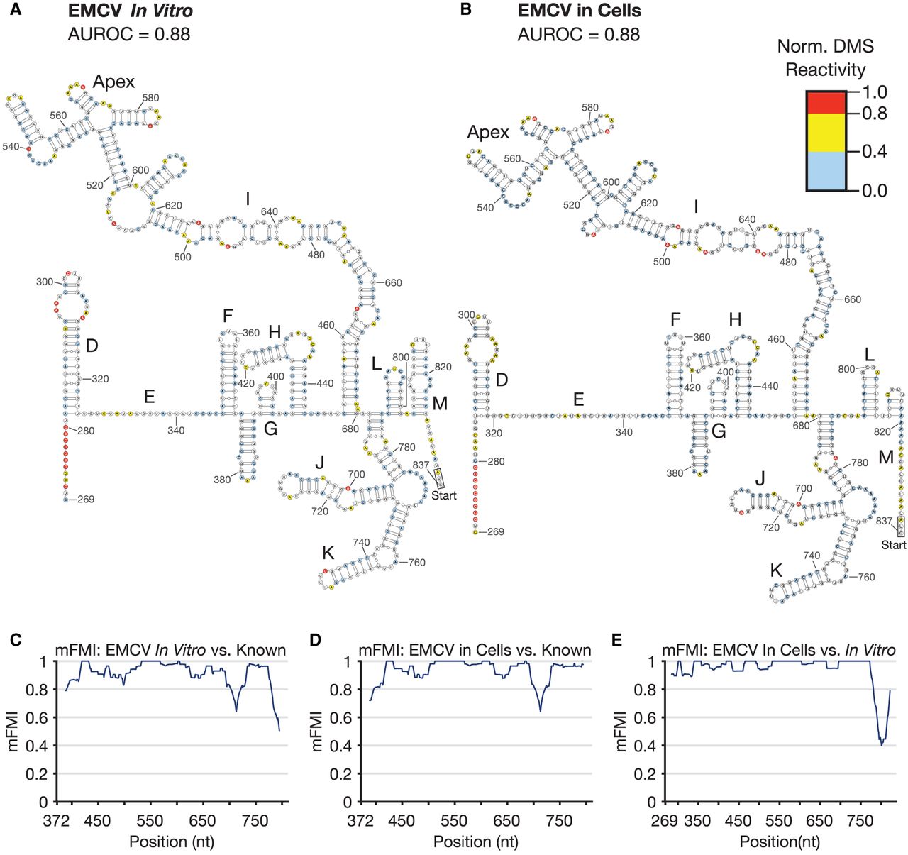
FIGURE 3.
Encephalomyocarditis virus (EMCV) IRES RNA secondary structure. (A) Secondary structure constrained by in vitro probing of the EMCV IRES (AUROC = 0.88). (B) The EMCV IRES secondary structure constrained by DMS-MaPseq, probed in HEK293T cells (AUROC = 0.88). (C) mFMI comparing EMCV probed in vitro (A) with the known structure reconstructed in Supplemental Figure S2B across their shared sequence range. (D) mFMI comparing EMCV in live cells (B) to the known structure. (E) mFMI between the cellular and in vitro models of the EMCV IRES (A vs. B).




