


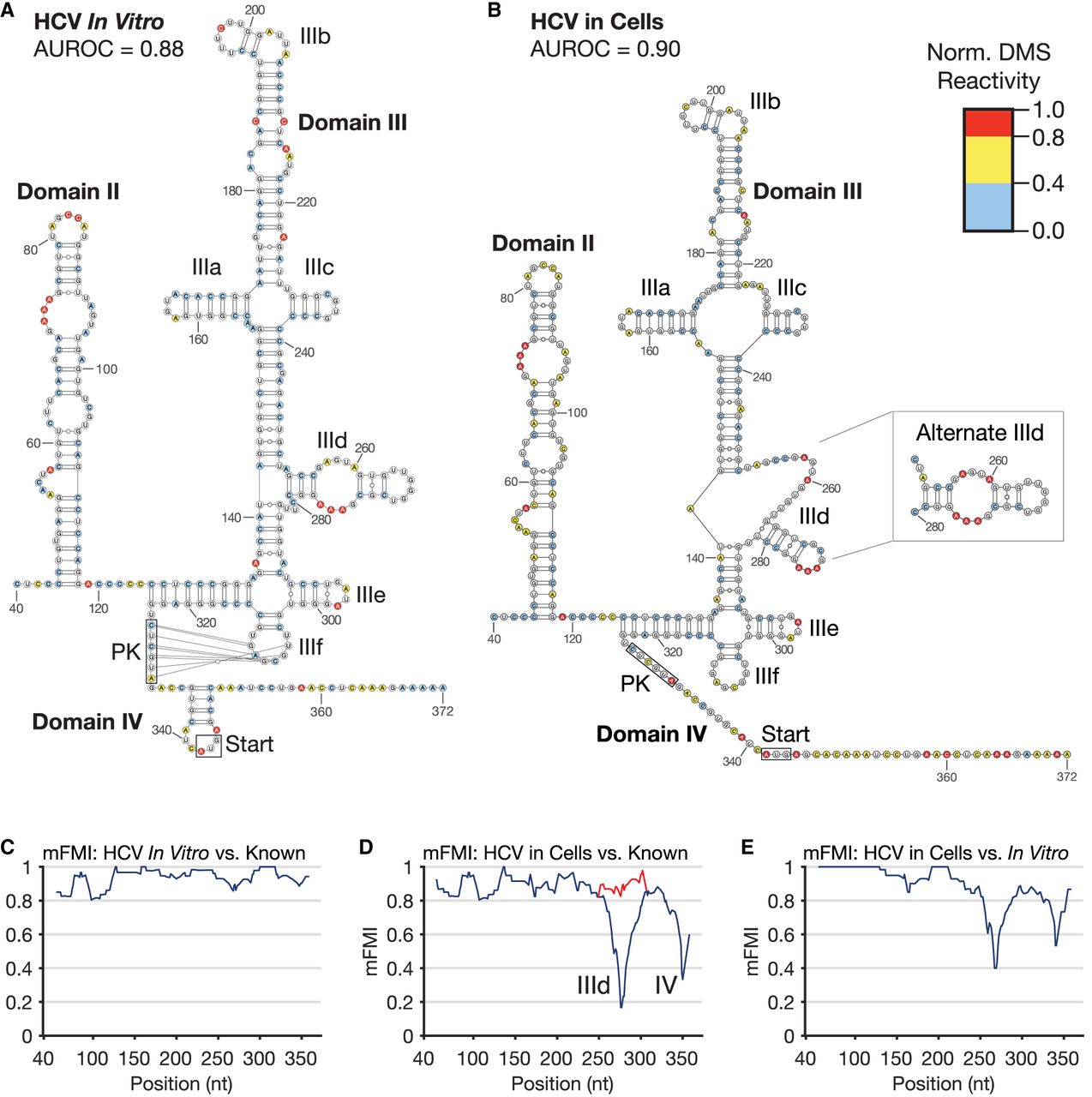
Hepatitis C virus (HCV) IRES RNA secondary structure. (A) The HCV IRES modeled from in vitro probing (AUROC = 0.88, see Materials and Methods). Normalized DMS reactivities calculated as described in Materials and Methods, and legend applies to all figures. White: no data (Gs and Us). Blue: shielded bases, 0.0–0.4. Yellow: bases of intermediate reactivity, 0.4–0.8. Red: highly reactive bases, 0.8–1.0. Domains of HCV are labeled consistently with previous reports. (B) The HCV 5′UTR IRES secondary structure constrained by DMS-MaPseq probing in HEK293T cells (AUROC = 0.90). At right, an alternative prediction of domain IIId, which more closely matches its known structure, is inlaid. (C) Modified Fowlkes–Mallows Index (mFMI, see Materials and Methods) quantitative structure comparison between the in vitro model and the known structure reconstructed in Supplemental Figure S2A. (D) mFMI comparing the cellular model and the same known structure. Notable areas of disagreement in base-pairing are labeled for domain IIId and domain IV. The red line corresponds to the mFMI of the alternative prediction inlaid at right of the structure. (E) mFMI between the cellular and in vitro models in A and B.




