


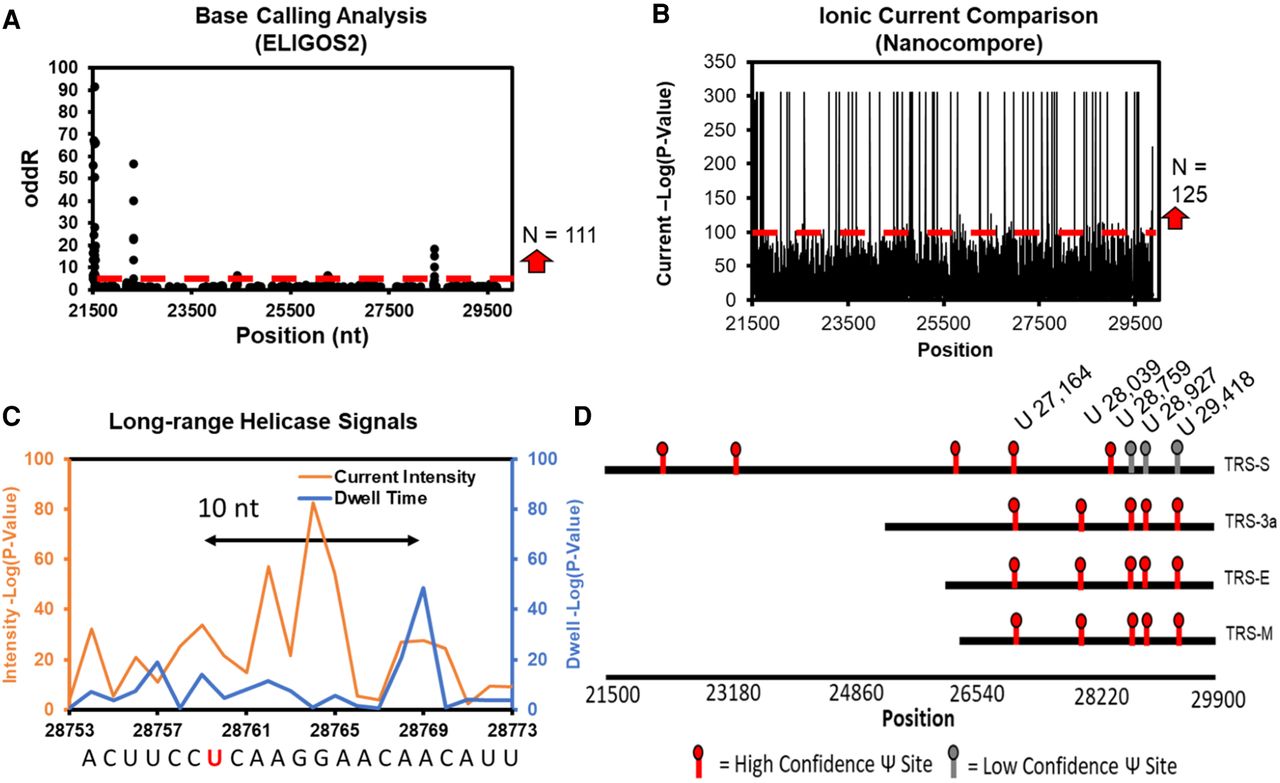
FIGURE 7.
Consensus analysis of DRS data for discovery of possible pseudouridinylation sites in SARS-CoV-2 gRNA. (A) ELIGOS2 base call analysis comparing viral RNA against a synthetic control without RNA modifications. (B) Nanocompore pairwise analysis of the ionic currents between the viral and synthetic RNAs. (C) Helicase dwell-time differences between the viral and synthetic RNA. (D) Sites identified to be pseudouridinylated based on the consensus analysis. The viral and synthetic data were obtained from publicly available sources (Kim et al. 2020; Miladi et al. 2020), and the figure is adapted from our prior work (Fleming et al. 2021).




