


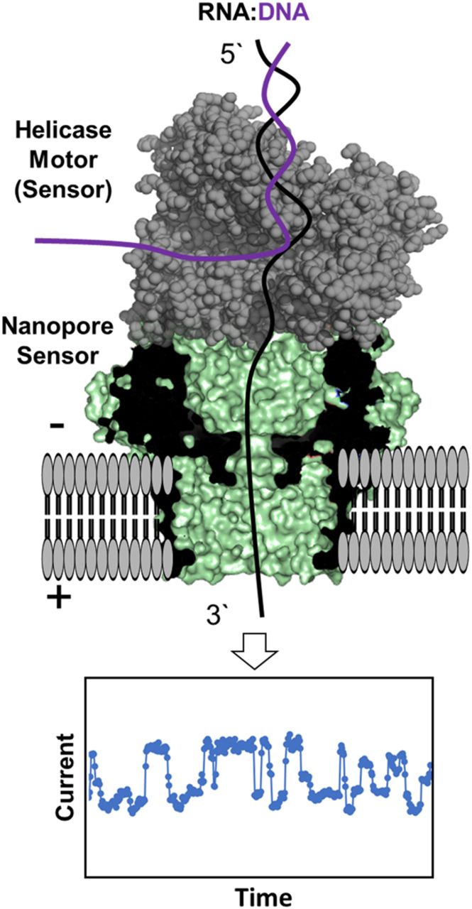
FIGURE 1.
Schematic of the nanopore sequencer illustrating the protein nanopore and helicase positioning to provide ionic current versus time traces as the RNA moves through the sensor. The figure for the nanopore sequencer was constructed from PDB 4UV3 and 2P6R (Büttner et al. 2007; Goyal et al. 2014).




