


FIGURE 8.
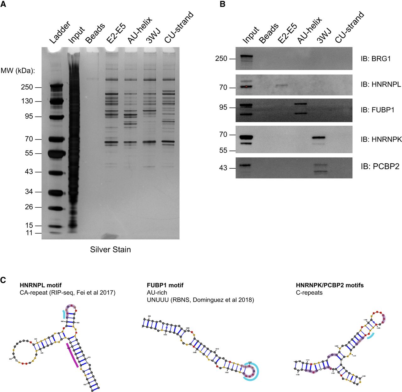
Identification of SChLAP1 substructure binding proteins. (A) Silver stain of pulldown eluates. Gel is representative of two independent experiments. (B) Western blot for BRG1, HNRNPL, FUBP1, HNRNPK, and PCBP2 in pulldown samples. Western blots are representative of two independent experiments. (C) Comparison of previously identified binding motifs to ΔSHAPE sites identified in our work. Structures of E2–E5 junction, AU-rich helix, and 3WJ with ΔSHAPE sites indicated are reprinted from Figure 4. Purple highlights represent motifs for HNRNPL, FUBP1, or HNRNPK/PCBP2.




