


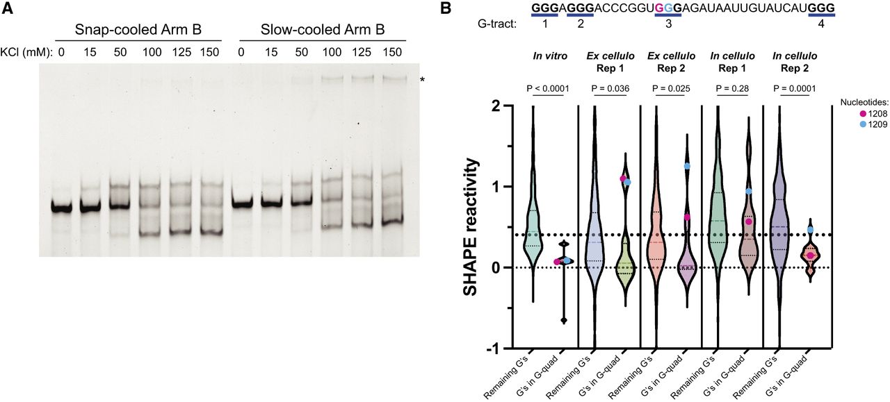
Analysis of G-quadruplex formation of Arm B by native gel electrophoresis and SHAPE. (A) Native gel electrophoresis of kit-purified Arm B in response to varying KCl concentrations and annealing conditions. Gel is representative of two biological replicates. (B, top) Sequence of predicted G-quadruplex in Arm B by QGRS Mapper (Kikin et al. 2006). G-tracts are numbered, and guanosines predicted to participate in G-quadruplex are in bold. (Bottom) Violin plots comparing the reactivities of guanosines in predicted G-quadruplex to the remaining guanosines in each SHAPE probing experiment reported herein. Reactivities below −1 or above 2 were not plotted here but were included in statistics. A line at SHAPE = 0.4 is shown for reference as SHAPE reactivities above this value are unfavorable for base-pairing (Hajdin et al. 2013). Significance was calculated using a Kolmogorov–Smirnov test for each experiment. Nucleotides 1208 and 1209 are indicated by color coding.




