


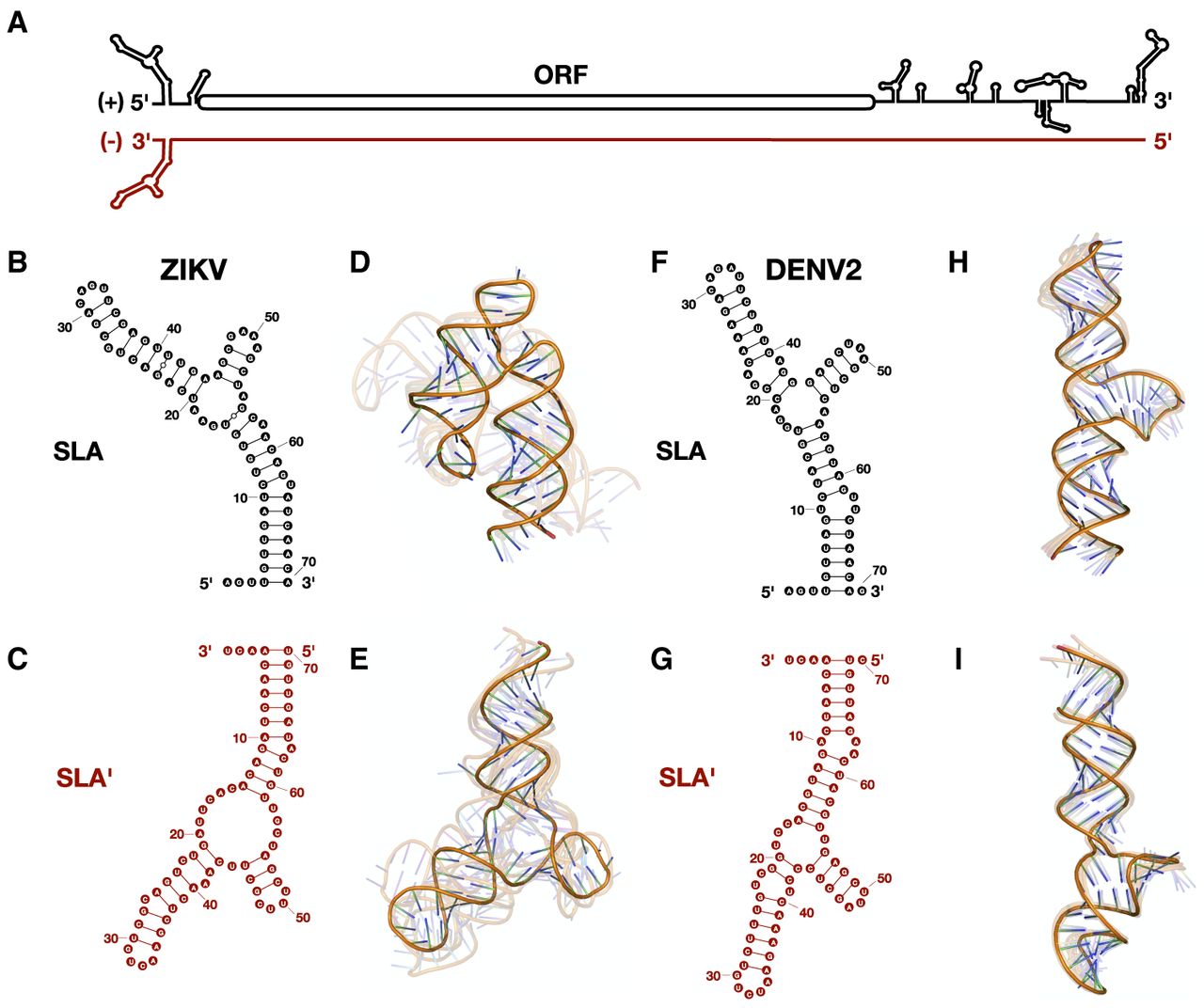
Zika virus (ZIKV) and Dengue virus 2 (DENV2) are predicted to form SLA′ at the 3′ terminus of their negative strands. (A) Cartoon diagram of the ZIKV positive-strand genomic RNA (black) and its complementary negative-strand intermediate (cayenne). Stem–loop A (SLA) is shown at the 5′ terminus of the positive strand, while the predicted SLA′ structure is shown at the 3′ terminus of the negative strand. Predicted secondary and tertiary structures of the first 70 nt of the 5′ terminus of the positive strand and the last 70 nt of the 3′ terminus of the negative strand of (B–E) ZIKV and (F–I) DENV2, respectively. Tick marks represent 10 nt intervals. The highest confidence tertiary structure prediction is fully opaque, while the next four highest-ranked predictions are translucent.




