


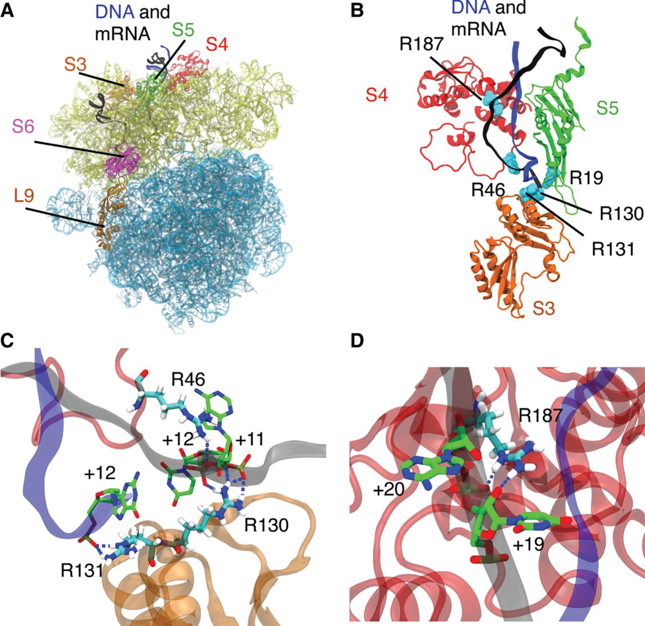
(A) The ribosome model used in MD simulations with water and ions omitted for clarity. The 50S subunit is shown in light blue, and the 30S subunit is shown in yellow. The ribosomal proteins S3, S4, and S5 are colored orange, red, and green, respectively. The mRNA is colored black, while the DNA 15 mer is colored blue. Proteins S6 and L9 used in the FRET experiments are shown in magenta and brown, respectively. (B) The position of arginines in the ribosomal proteins S3, S4, and S5 that were found to maintain hydrogen bonds with either DNA or mRNA residues 50–70 in more than 50% of the trajectory frames for the +11 construct. The arginines are shown in cyan, and DNA and mRNA are shown in blue and black, respectively. Proteins S3, S4, and S5 are colored as in A. (C) Selected snapshot from our simulations illustrates hydrogen bonding of protein residues R130 and R131 from S3 and R46 from S4 with the DNA–mRNA strand. The backbone colors for DNA, mRNA, S3, and S4 are the same as in A. The protein and nucleic residues directly involved in hydrogen bonding are shown in licorice representation. For easier visualization, the carbon atoms are colored cyan for arginine residues and green for nucleic acid residues. Hydrogen atoms are shown in white and are omitted for nucleic acid residues for clarity. For all residues, oxygen atoms are colored red, and nitrogen atoms are blue. Hydrogen bonds are visualized as dotted blue lines. (D) Selected snapshot from our simulations illustrates hydrogen bonding between R187 from S4 and mRNA residues +19 and +20 in the +11 system. The colors and representations are the same as in C.




