


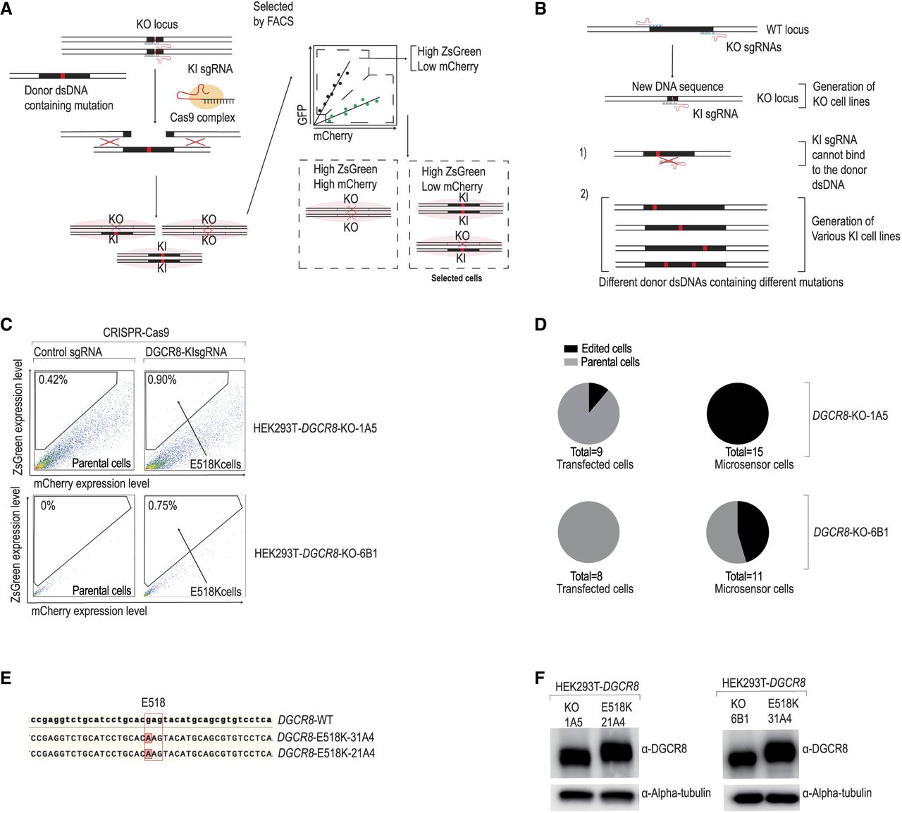
Using Microsensor-assisted CRISPR-Cas9 editing for precise DGCR8 mutation. (A) Workflow for generating DGCR8 mutant cells. The DGCR8-KO cells, which had a specific DNA region deleted containing the mutation of interest, were transfected with KI (knockin) sgRNA, Cas9 plasmids, and the Microsensor system, along with donor dsDNA harboring a mutation site. FACS was used to first select transfected cells based on the ZsGreen signal and then to identify potential mutant cells showing a high ZsGreen to mCherry ratio, which indicated DGCR8-KI cells. The cells were sorted individually and cultured in wells for 2–3 weeks for further analysis. (B) Advantages of generating mutant DGCR8 cells using a two-step approach. The KO cells generate a new DNA sequence at the mutation site, distinct from that of wild-type cells, allowing for the specific design of KI sgRNA that targets only this genomic DNA, and not the donor DNAs in knock-in experiments. The KO step produces various KO cells, serving as a platform that can be targeted by any donor DNA containing different mutations. (C) FACS results of DGCR8-KI experiments. The KI experiments conducted on two different DGCR8-KO cells, 1A5 and 6B1. The FACS analysis of the DGCR8-KI cells displayed the ZsGreen level on the y-axis and the mCherry level on the x-axis. The marked area highlighted the cells potentially modified through the KI experiments, showing a high ratio of ZsGreen to mCherry. (D) Pie chart of edited cells. These pie charts illustrate the proportion of cells containing the KI-band, as identified from the PCR experiments described in Materials and Methods. (E) Sequencing analysis of DGCR8-KI cells. DNA regions harboring mutations from DGCR8-KI cells (clones 21A4 and 31A4) were amplified via PCR, and the amplified DNA was sequenced directly using Sanger sequencing. The mutated nucleotides are highlighted. (F) Western blot analysis. This analysis was performed on DGCR8-KO cells (1A5 and 6B1) and DGCR8-KI cells (21A4 and 31A4).




