


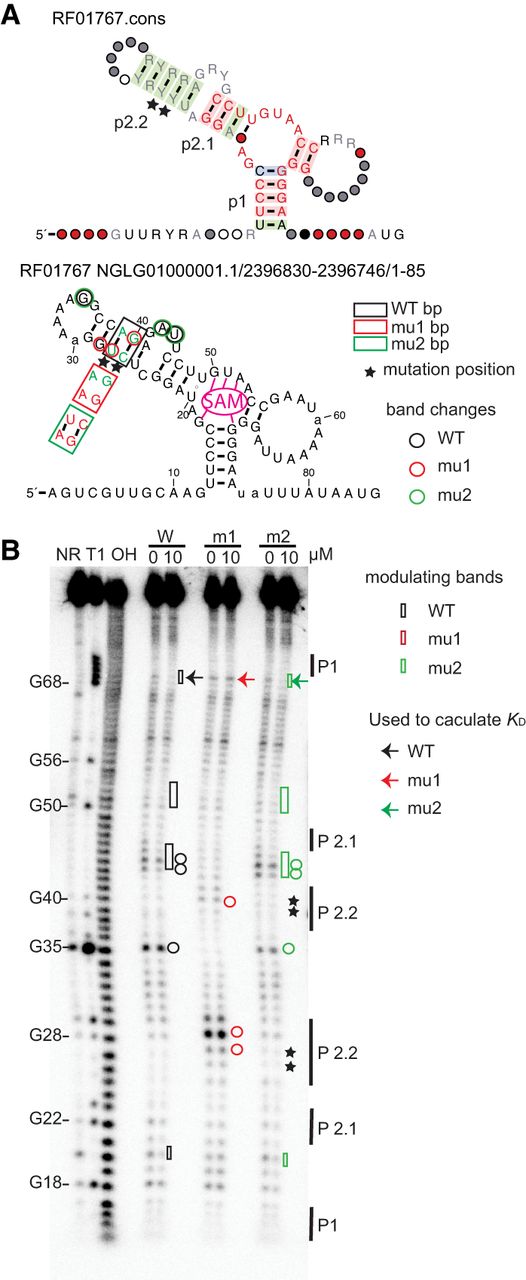
SAM-III riboswitch. (A, top) The consensus structure of the SAM-III riboswitch, see legend from Figure 2A. (A, bottom) The secondary structure of the sequence from Enterococcus sp. 3H8_DIV0648 used in in-line probing assay. The green nucleotides represent wild-type nucleotides, while the red nucleotides indicate the mutated nucleotide. The different colored circles show the structural changes among three different constructs, mapped from the gel result in B. (B) In-line probing gel patterns from the three different constructs of SAM-III and the modulation changes in the presence of SAM. The different colored rectangles indicate ligand-induced modulation. The arrows mark the positions used to calculate the KD. The stars mark the mutation positions. The black solid lines mark the stems of the secondary structure. The circles indicate the strongest bands corresponding to structural change among the wild-type, mu1, and mu2 constructs.




