


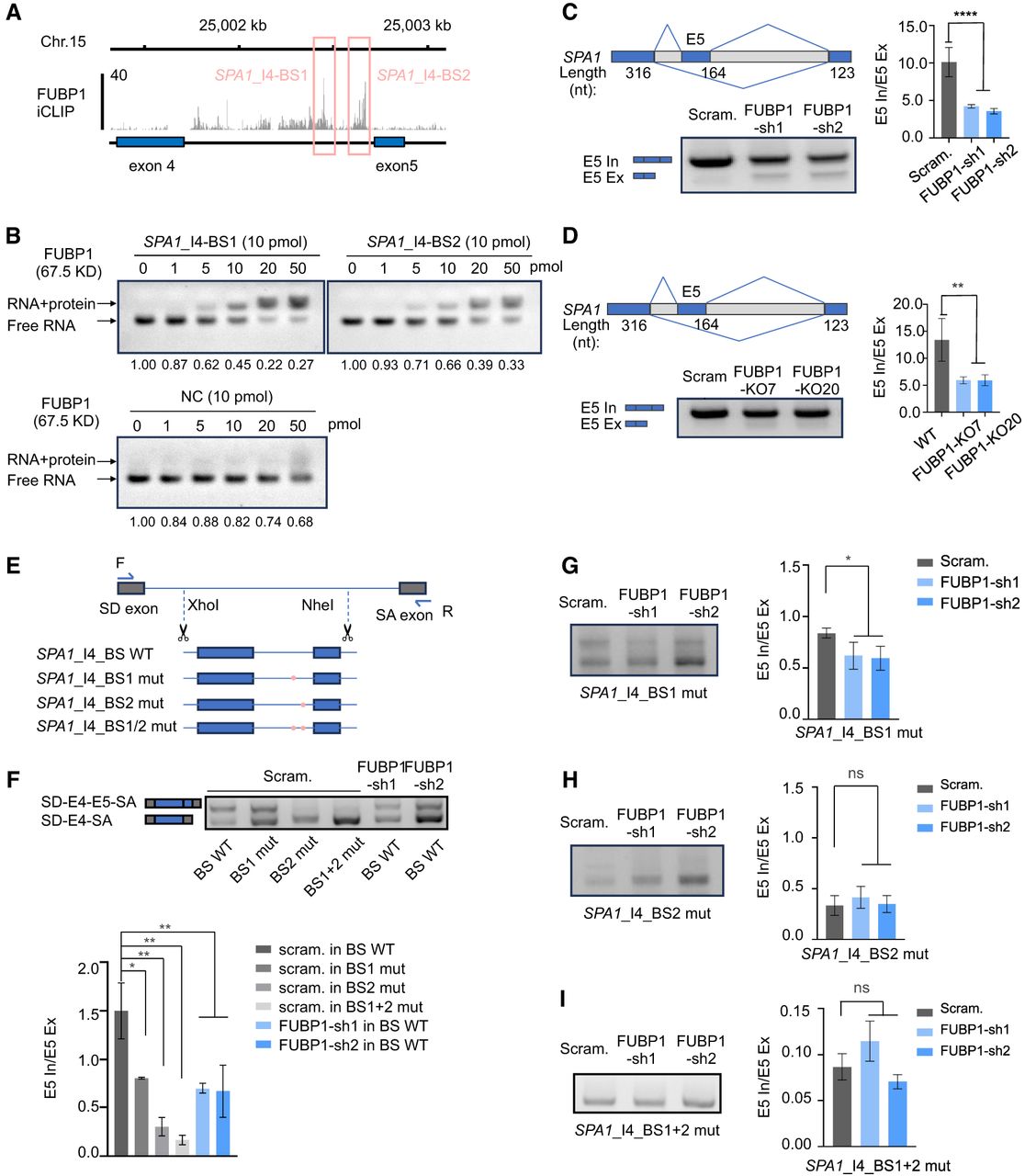
FUBP1 regulates splicing of SPA1 via the U-rich sequence. (A) FUBP1 iCLIP-seq results (GEO: GSE220184) (Ebersberger et al. 2023) showing strong binding to the 3′ of the SPA1 intron 4. The pink boxes refer to the FUBP1 binding sites named SPA1-I4-BS1 and SPA1-I4-BS2. (B) FUBP1 binds the SPA1-I4-BS1 and SPA1-I4-BS2, revealed by EMSA. (C) FUBP1 affects the splicing of SPA1 in PA1 cells. (Upper left) Schematic of SPA1 splicing. The length of SPA1 exons is indicated. (Lower left) Agarose gel image showing that loss of FUBP1 results in SPA1 exon 5 skipping. (D) FUBP1 affects the splicing of SPA1 in H9 cells. (Upper left) Schematic of SPA1 splicing. The length of SPA1 exons is indicated. (Lower left) Agarose gel image showing that loss of FUBP1 results in SPA1 exon 5 skipping. (E) Schematic of the minigene assay design. SPA1_I4_BS_WT, SPA1_I4_BS1 mutation, SPA1_I4_BS2 mutation or SPA1_I4_BS1/2 double mutant is inserted in the plasmids. (F) Expressing the minigene containing the BS1 and BS2 single or double mutation in the scramble PA1 cells or expressing the minigene containing BS_WT in the FUBP1 knockdown PA1 cells results in increased exon 5 skipping of SPA1, revealed by RT-PCR. (G) Knockdown of FUBP1 results in the increased exon 5 skipping of SPA1_I4_BS1 mutant, revealed by RT-PCR. (H) Knockdown of FUBP1 barely affects the splicing of SPA1_I4_BS2 mutant, revealed by RT-PCR. (I) Knockdown of FUBP1 barely affects the splicing of SPA1_I4_BS1 and SPA1_I4_BS2 double mutant, revealed by RT-PCR. (C, D, F–I). Statistics of three biological repeats of the gels are shown.




