


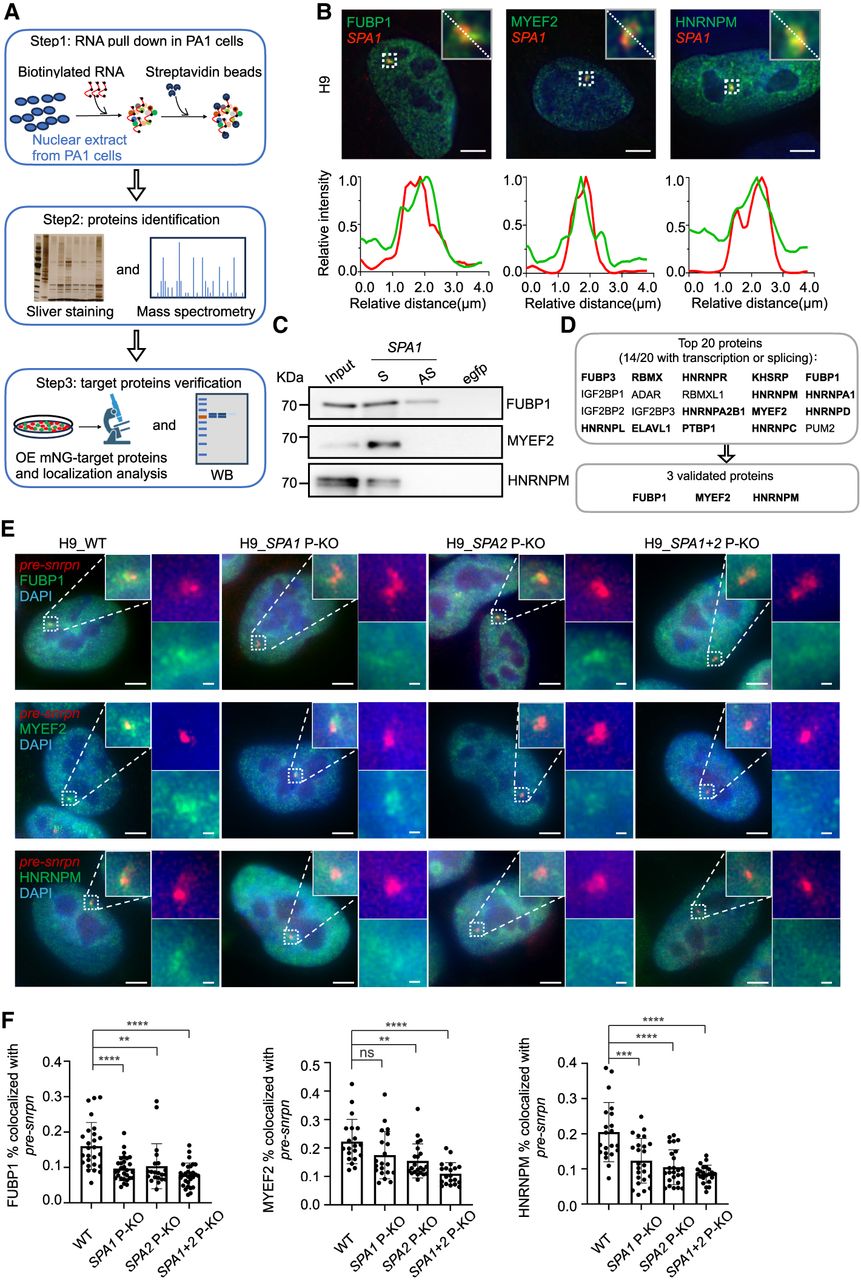
SPAs enrich FUBP1, MYEF2, and HNRNPM in PWS bodies in H9 cells. (A) Schematic of the workflow for identifying the SPA1-interacting proteins. The nuclear extract from PA1 cells is used for the RNA pull-down assay (step 1). The pull-down quality is confirmed by silver staining, and the interacting proteins are identified by mass spectrometry (step 2). The interacting proteins are further validated by imaging and western blotting (step 3). (B) Colocalization of SPA1 with FUBP1 (left), MYEF2 (middle), and HNRNPM (right). (Top) Representative images showing the colocalization. (Bottom) Line scans of the relative fluorescence intensity from the representative images above. Scale bar, 5 μm. (C) Western blotting showing the interaction between SPA1 and FUBP1, MYEF2, or HNRNPM in H9 cells. Proteins are pulled down by biotin-sense (S) SPA1, biotin-antisense (AS) SPA1, and biotin-egfp from H9 cells. (D) Top 20 SPA1-interacting proteins identified from (A). Fourteen of them are associated with transcription or splicing regulating (marked in bold). Among them, FUBP1, MYEF2, and HNRNPM are further validated. (E) The representative images showing that paternal depletion of SPA1, SPA2, or SPA1+2 reduces the enrichment of FUBP1, MYEF2, and HNRNPM in the PWS bodies in H9 cells. Scale bar, 5 μm (uncropped) and 500 nm (cropped). pre-snrpn is stained as the marker of PWS bodies. (F) Percentage of the PWS body localized FUBP1, MYEF2, and HNRNPM evidenced by colocalization with pre-snrpn in H9 cells. For evaluating PWS body localized FUBP1, WT (n = 26), SPA1 P-KO (n = 29), SPA2 P-KO (n = 21), and SPA1+2 P-KO (n = 29) cells are used. For evaluating PWS body localized MYEF2, WT (n = 20), SPA1 P-KO (n = 20), SPA2 P-KO (n = 22), and SPA1+2 P-KO (n = 20) cells are used. For evaluating PWS body localized HNRNPM, WT (n = 21), SPA1 P-KO (n = 24), SPA2 P-KO (n = 25), and SPA1+2 P-KO (n = 22) cells are used.




