


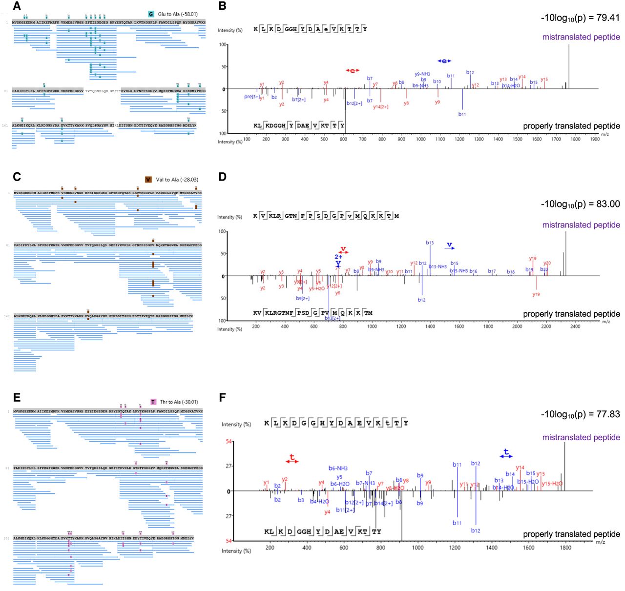
tRNA-dependent misincorporation of Ala detected by mass spectrometry. Tandem mass spectrometry analysis revealed Ala misincorporation in mCherry isolated from HEK 293T cells expressing (A, B) tRNAAlaCUC(Glu), (C, D) tRNAAlaUAC(Val), and (E, F) tRNAAlaAGU(Thr) at (A, B) Glu, (C, D) Val, and (E, F) Thr codons, respectively. (A, C, E) Misincorporation events in mCherry are depicted in peptide coverage maps alongside (B, D, F) example mirror spectra comparing mistranslated peptides (top) to the properly translated matching peptide (bottom). Colored boxed symbols above the peptide sequence annotate the indicated mistranslated residues identified with minimal ion intensities of ≥2%. The data are based on n = 3 biological replicates.




