


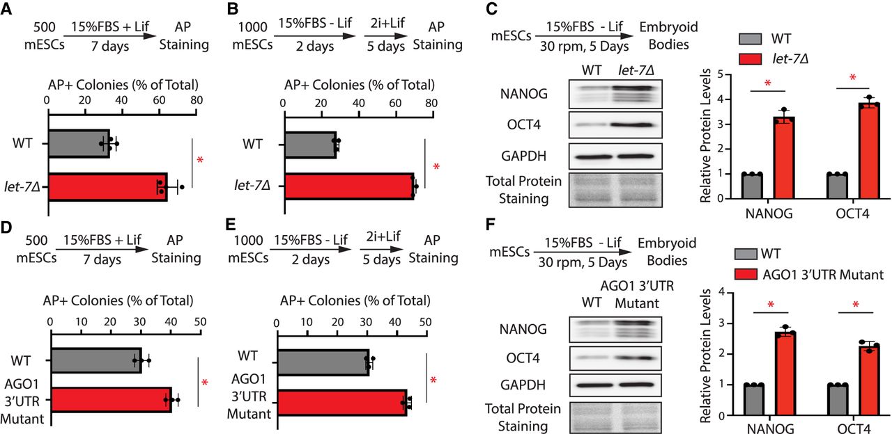
let-7-mediated repression of AGO1 is important for mESC differentiation. (A) Colony formation assay for the WT and the let-7Δ mESCs. (B) Exit pluripotency assay for the WT and the let-7Δ mESCs. (C) Western blotting in the differentiating WT and the let-7Δ mESCs. (D) Colony formation assay for the WT and the AGO1 3′-UTR mutant mESCs. (E) Exit pluripotency assay for the WT and the AGO1 3′-UTR mutant mESCs. (F) Western blotting in the differentiating WT and AGO1 3′-UTR mutant mESCs. Data information: In A–B and D–E, the colony morphology and AP intensity were evaluated via microscopy. In total, 100–200 colonies were examined each time to determine the percentage of undifferentiated colonies. In C and F, representative western blots are shown. All the quantifications represent the means (±SD) of three independent experiments. GAPDH levels were used for normalization in calculating the relative protein levels. One-way ANOVA was used to determine the significance of the difference, (*) P < 0.05.




