


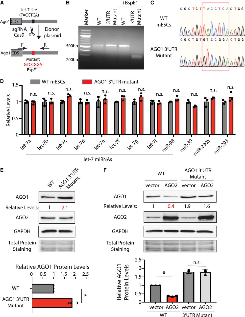
AGO1 is regulated by let-7 miRNAs in mESCs. (A) Mutating the let-7 miRNA binding site in AGO1 mRNA 3′ UTR via genome editing. (B) Genotyping of the AGO1 3′-UTR mutant. The PCR was performed using the oligos (F and R) indicated in A. (C) Sanger sequencing to verify the biallelic mutation of the let-7 miRNA-binding site in AGO1 mRNA 3′ UTR. The red box indicates the let-7 miRNA-binding site. (D) qRT-PCR quantification of miRNAs in the WT and the AGO1 3′-UTR mutant mESCs. U6 RNA was used for normalization. The results represent the means (±SD) of three independent experiments. One-way ANOVA was used to determine the significance of the difference, (n.s.) not significant (P > 0.05). (E) Western blot in the WT and the AGO1 3′-UTR mutant mESCs cultured in the 15% FBS + Lif medium. (F) Western blotting in the WT and the AGO1 3′-UTR mutant mESCs expressing either a vector or AGO2. Data information: In E and F, representative western blots are shown. All the quantifications represent the means (±SD) of three independent experiments. GAPDH levels were used for normalization in calculating the relative protein levels. One-way ANOVA was used to determine the significance of the difference, (*) P < 0.05, (n.s.) not significant (P > 0.05).




