


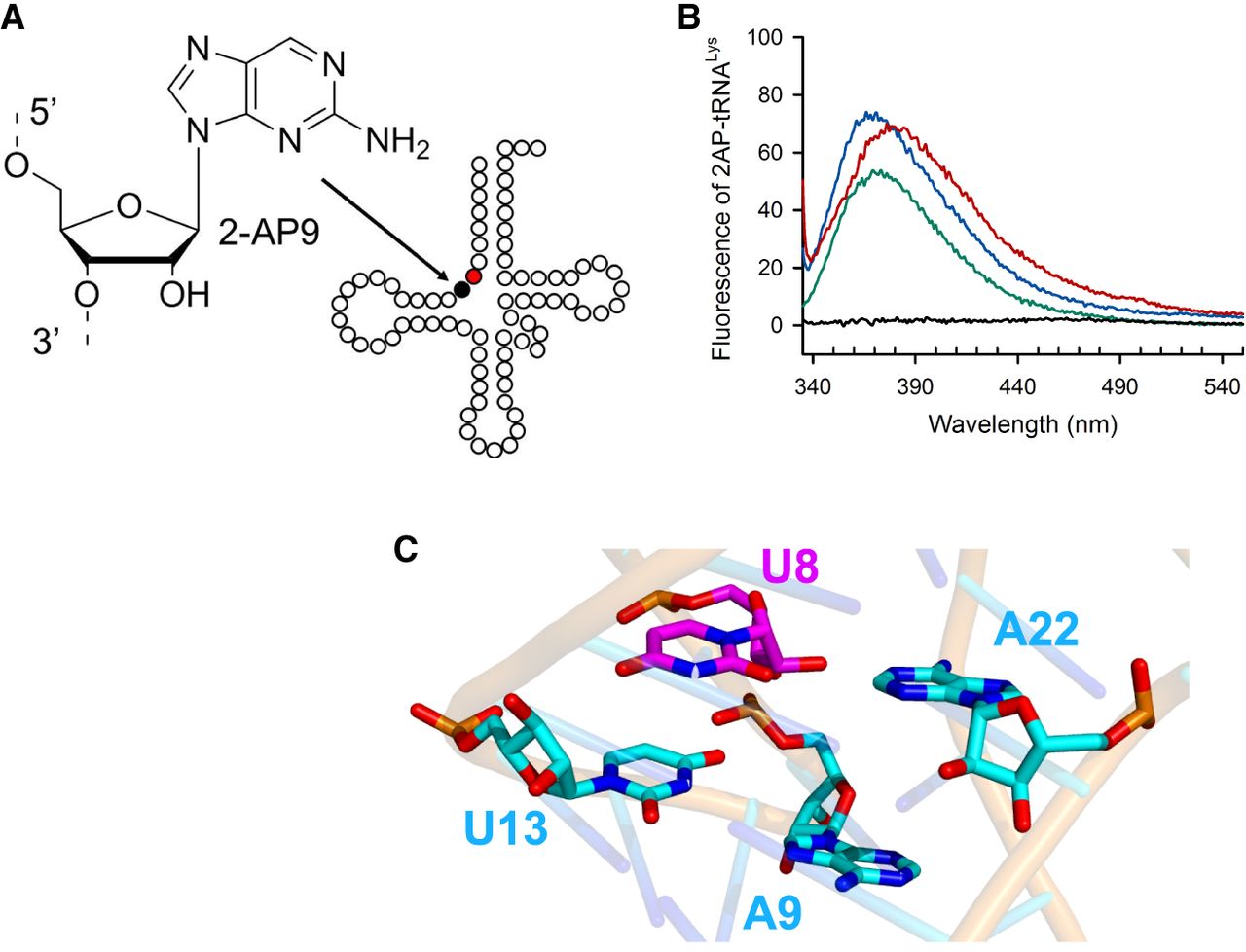
FIGURE 3.
Sensitivity of the [4Fe–4S] cluster of MmTtuI to the environment of the U8 target base in MmtRNALys. (A) Chemical structure of 2-aminopurine (2AP) and its position (in black) in 2AP9-MmtRNALys, adjacent to the target base U8 (in red). (B) Fluorescence spectra obtained at λex = 320 nm of 2AP9-MmtRNALys (0.5 µM). 2AP9-MmtRNALys alone (in green), in the presence of apo-MmTtuI (2 µM) + 3 mM AMP-CPP (in blue) or in the presence of holo-MmTtuI (2 µM) + 3 mM AMP-CPP (in red). Apo-MmTtuI in the absence of 2AP9-MmtRNALys (in black). (C) Region surrounding U8 in the AlphaFold3 model of MmtRNALys (pTM score of 0.44). The target base U8 is shown in magenta.




