


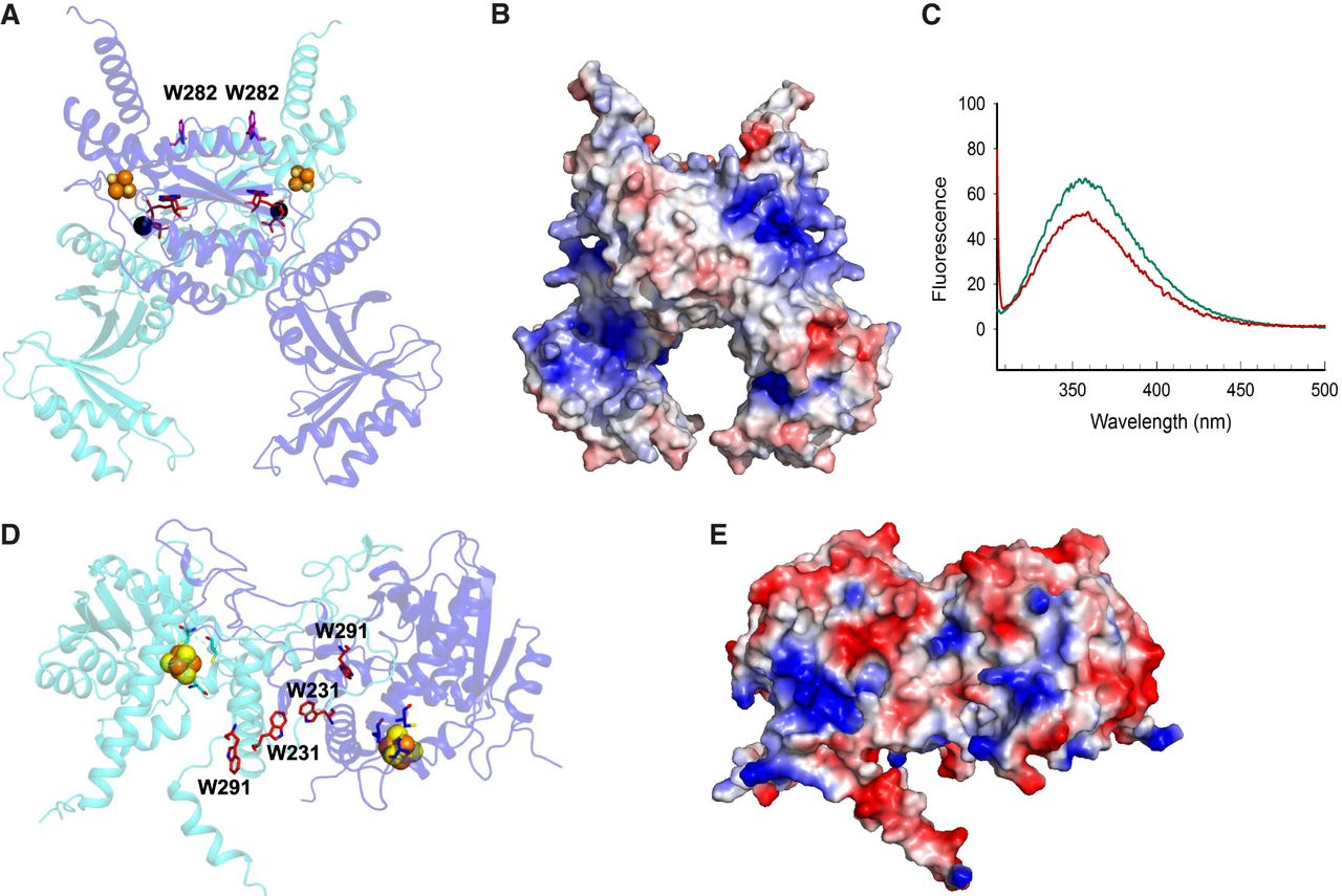
Environment of tryptophan residues in MmTtul and EcTtcA. (A) AlphaFold2 model of the holo-MmTtuI dimer in complex with ATP and Mg2+. One monomer is colored in cyan and the other in blue. Residues W282 and ATP are depicted as sticks (in magenta and red, respectively), while the [4Fe–4S] cluster and Mg2+ ions are shown in sphere representation with sulfur atoms colored in yellow, iron atoms in orange, and Mg atoms in black. (B) Electrostatic surface of the holo-MmTtuI dimer (negative charge in red, positive charge in blue). (C) Fluorescence spectra obtained at λex = 295 nm of native apo-MmTtuI (in green) and after denaturation with 8 M urea (in red). (D) AlphaFold2 model of the holo-EcTtcA dimer. One monomer is colored in cyan and the other in blue. Residues W231 and W291 are depicted as red sticks, while the [4Fe–4S] cluster is shown in sphere representation. (E) Electrostatic surface of the holo-EcTtcA dimer.




