


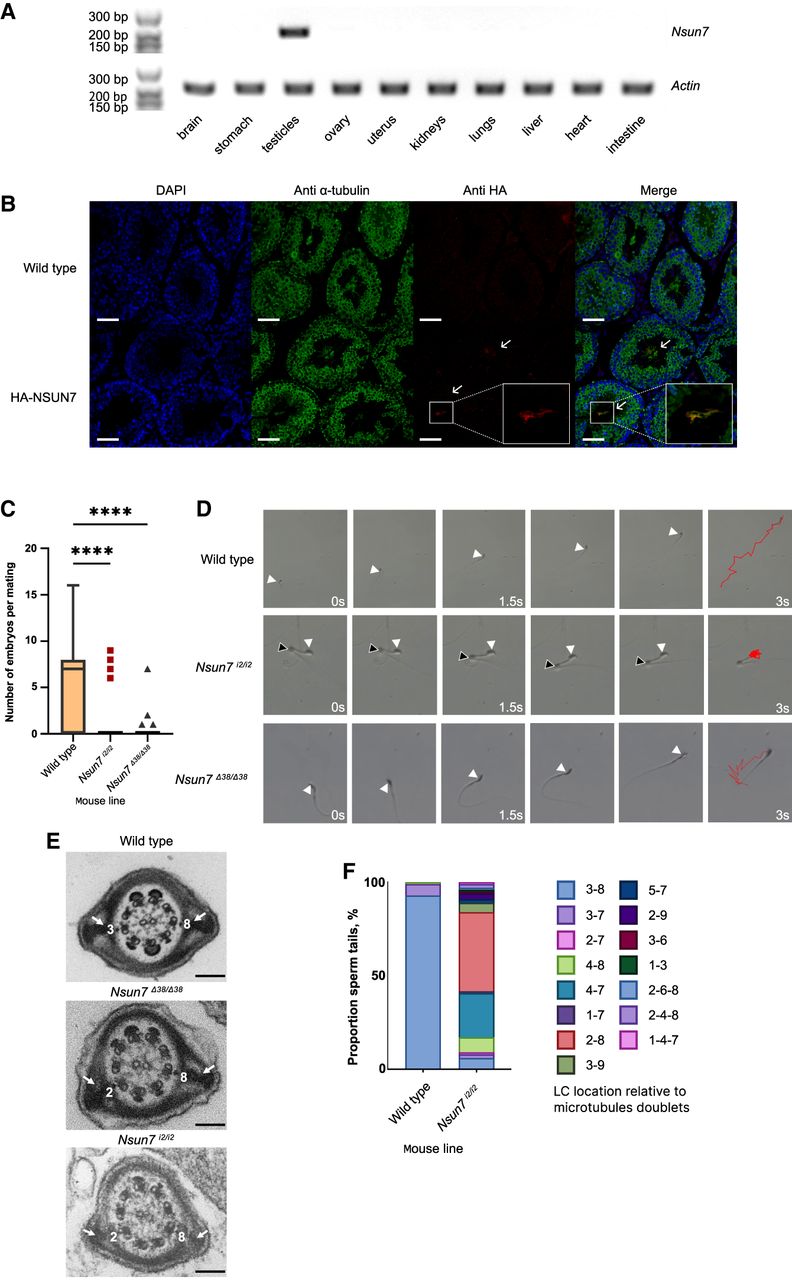
Nsun7 is expressed in testes. (A) RT-PCR analysis of Nsun7 gene expression in mouse organs. ActB gene is used as a positive control. (B) Immunostaining of testes cross sections of the wild-type and HA-Nsun7 mice. White bars = 100 μm. Arrows indicate HA-NSUN7 localization in flagella of elongated spermatids. The colocalization of HA-NSUN7 and α-tubulin is shown in the frames on a higher magnification. (C) Decrease of fertility of Nsun7i2/i2 (n = 21) and Nsun7Δ38/Δ38 (n = 18) mouse lines relative to the WT (n = 47). For each mouse line, the number of embryos was analyzed 10 dpc. ANOVA test result: (****) P < 0.0001. (D) Motion tracks of spermatozoa of the WT (top panel), Nsun7i2/i2 (middle panel), and Nsun7Δ38/Δ38 (bottom panel) mice. Red lines show the motion tracks of spermatozoa, white arrowheads indicate head position, black arrowheads—cytoplasmic bugling position. (E) Electron microscopy of spermatozoid tails of the WT (top panel), Nsun7i2/i2 (middle panel), and Nsun7Δ38/Δ38 (bottom panel) mice. White arrows indicate LC. Black bars = 100 nm. (F) Distribution of LC position in the WT (n = 3 mice) and Nsun7i2/i2 (n = 3 mice).




