


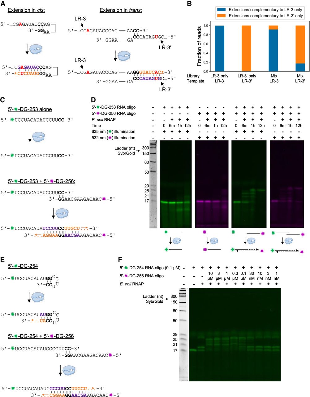
RNA-templated RNA extension in cis and in trans. (A) Schematic of experiment to test cis versus trans nature of the extension of LR-3. The single base difference between LR-3 and LR-3′ is bolded in red, and RNAP is in blue; otherwise, colors for the extension sequences are as in Figure 3B. (B) Oligonucleotides LR-3, LR-3′, and an equimolar mixture of LR-3 and LR-3′ were extended with E. coli RNAP, and the extension products were sequenced. For each library (LR-3, LR-3′, and mixture) and for each template (LR-3 and LR-3′), the fraction of extensions complementary to LR-3 alone (blue) or LR-3′ alone (orange) was counted. Extensions complementary to both LR-3 and LR-3′ were not counted here. (C) Schematic of experiment to test the possibility of trans RNA-templated RNA extension. (D) Trans extension of two RNA oligonucleotide templates. White space indicates gel splicing, and RNAP is in blue. (E) Schematic of experiment to attempt to assess cis and trans RNA-templated RNA extension in the same reaction. (F) Analysis of cis and trans RNA-templated RNA extension in the same reaction. Note that since DG-254 concentration was reduced from our default of 1 to 0.1 μM, RNAP concentration was reduced from 0.85 μM to 85 nM. Extension was for 1 h. In C and E, colors for the extension sequences are as in Figure 3B, and RNAP is in blue. In D and F, RNAP reactions were analyzed crude on 15% gels with gel protocol 2 (see Materials and Methods). Panels pseudocolored green reflect 635 nm illumination and visualization of TYE 655 (green star), and panels pseudocolored red reflect 532 nm illumination and visualization of Cy3 (red star). In all cases, all fluorophores were imaged first; the gel was then stained with SybrGold and reimaged (to visualize the ladder). The diffuse stain at ∼30 nt with 635 nm illumination is xylene cyanol.




