


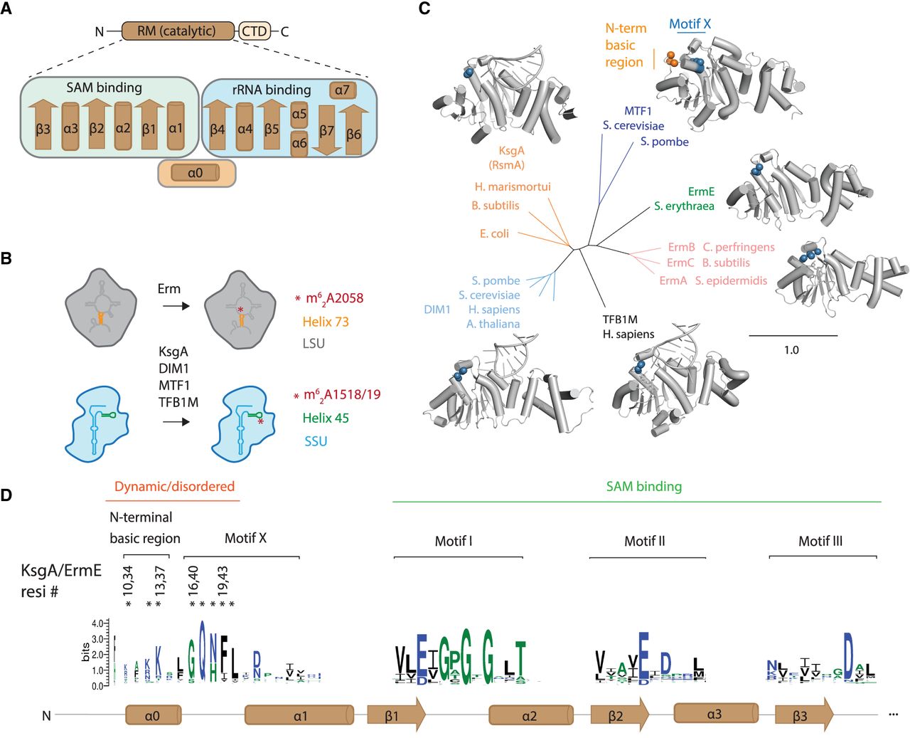
The structure and activity of RRAD family members. (A) RRAD enzymes consist of a Rossmann-like methyltransferase (RM) catalytic domain and a C-terminal domain. The secondary structure of the RM domain is shown indicating that α1–β3 are predominantly involved in SAM while β4–β7 are involved in rRNA binding. (B) RRAD family members dimethylate rRNA of either the large ribosomal subunit (LSU) or small ribosomal subunit (SSU). (C) Experimentally determined structures are available for each RRAD family member in some cases bound to rRNA (MTF1, PDB code 1i4w; ErmE, 6nvm; ErmC, 1qao; TFB1M, 6aax; DIM1, 7wtm; KsgA, 7o5h). An N-terminal basic region (orange) is disordered in these structures (except for MTF1), while the adjacent motif X (blue) is partially ordered. (D) The conservation of RRAD family members at key motifs throughout the α0–β3 region of the protein family is shown as a sequence logo. Residues in the N-terminal basic region and motif X investigated by site-directed mutagenesis in this study are marked with an asterisk (*).




