


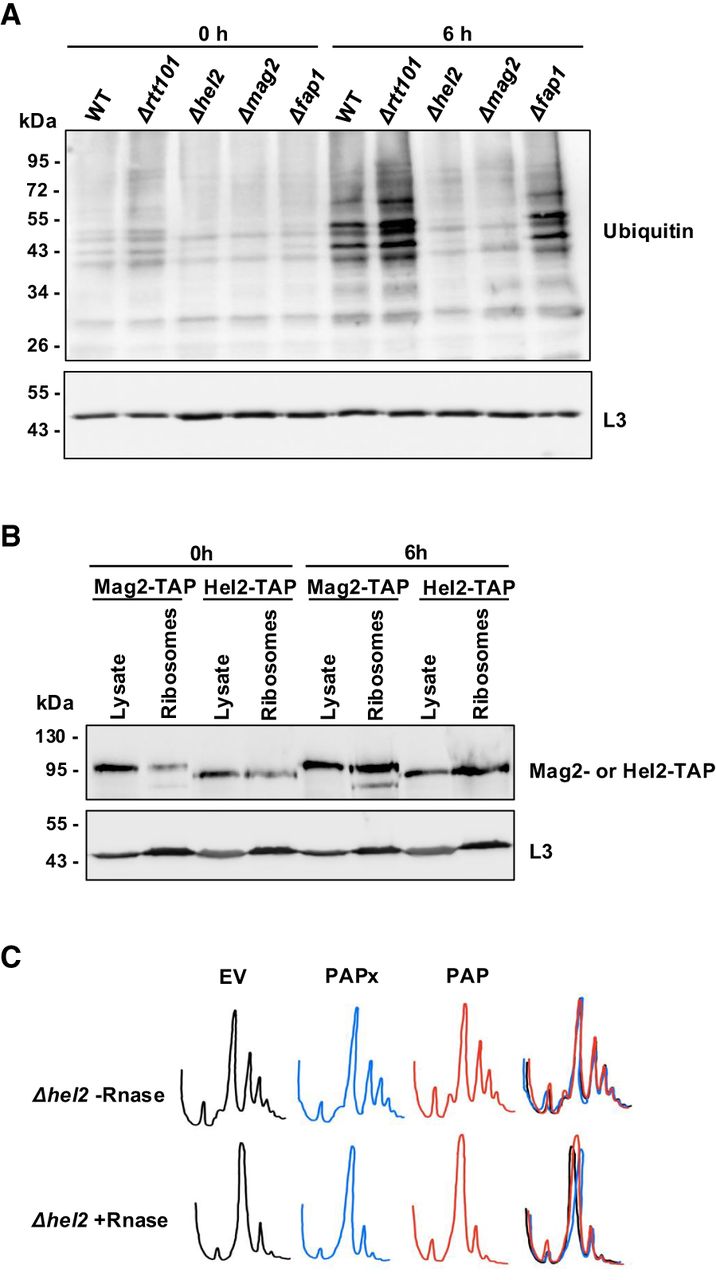
PAP activates 18S NRD without causing ribosome collisions. (A) Immunoblot illustrating ubiquitination of ribosomes from WT, Δrtt101, Δhel2, Δmag2, and Δfap1 BY4741 cells transformed with a cDNA encoding PAP at 0 and 6 h postinduction with 2% galactose. Ribosomes (20 µg) were separated through 12% SDS PAGE, transferred to nitrocellulose, and probed with a ubiquitin antibody (1:1000). A monoclonal antibody against RpL3 (43 kDa; 1:5000) was used as a loading control. The blots shown are representative of three independent biological replicates (n = 3). (B) Immunoblot of TAP-tagged Hel2 (93 kDa) and Mag2 (96 kDa), in total cell lysates and ribosomes isolated from Hel2-TAP and Mag2-TAP tagged BY4741 cells transformed with a cDNA encoding PAP at 0 and 6 h postinduction with 2% galactose. Total cell lysate and ribosomes (40 µg) were separated through 12% SDS PAGE, transferred to nitrocellulose, and probed with a polyclonal TAP (1:1000) antibody. A monoclonal antibody against RpL3 (43 kDa; 1:5000) was used as a loading control. The blots shown are representative of three independent biological replicates (n = 3). (C) Evaluation of disome presence in sucrose gradient profiles of PAP-expressing cells. Representative sucrose gradient profiles of Δhel2 BY4741 cells transformed with cDNAs encoding PAP, PAPx, and EV following induction with 2% galactose for 6 h. Lysates were treated with or without RNase-I before separation on a 20%–50% sucrose gradient. Absorbance of collected fractions was measured at A260.




