


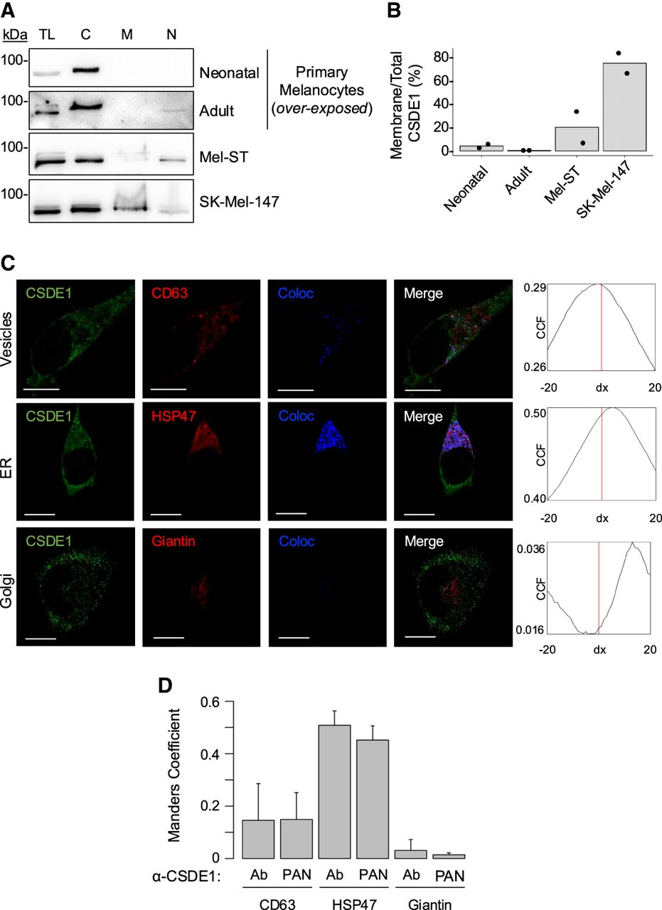
CSDE1 localizes with membrane compartments in melanoma cells. (A) Fractionation of CSDE1 in melanocytes (neonatal or adult), immortalized Mel-ST cells, and metastatic melanoma cells. (TL) Total lysate, (C) cytosol, (M) membranes, (N) nucleus. (B) Quantification of CSDE1 levels in membranes (n = 2). CSDE1 amounts in the membrane and total fractions were corrected by an internal control (HSP47 levels) and expressed as percentage of total CSDE1. (C) CSDE1 colocalization with membrane compartments assessed by confocal microscopy. Typical images obtained with the Abcam (ab201688) αCSDE1 antibody are shown. Scale bar, 10 µm. The Van Steensel cross-correlation coefficient (CCF) is shown on the right. (D) The Manders coefficient for colocalization of CSDE1 and the indicated markers using both the Abcam (ab201688) and “PAN” αCSDE1 antibodies. Bars represent the average of five cells, and error bars represent standard deviation.




