


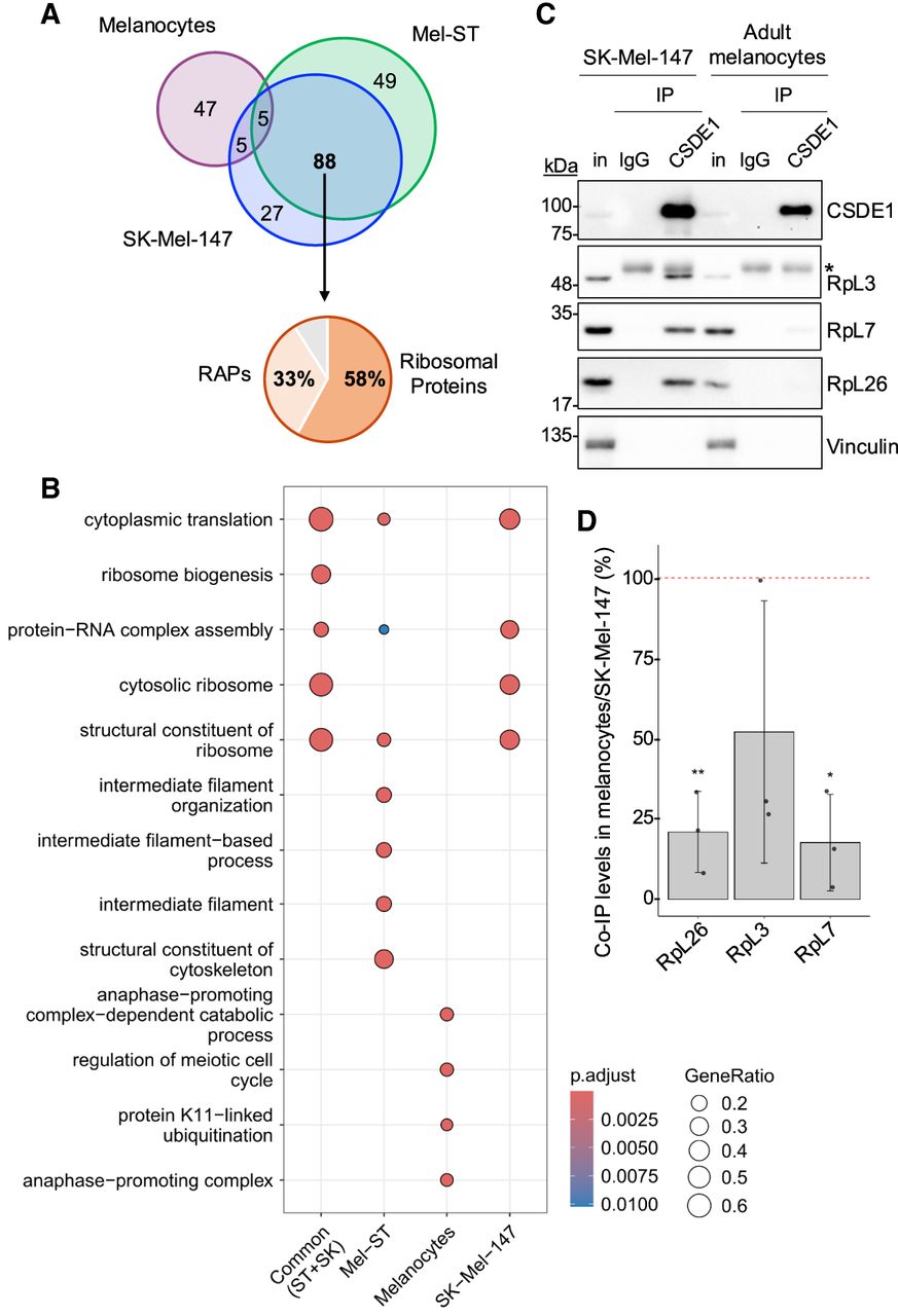
CSDE1 interacts with ribosomes in melanoma cells. (A) Venn diagram showing the overlap of CSDE1 interactors identified in cells of melanocytic origin (n = 3 per cell type). RAPs, ribosome associated proteins. (B) GO term categories of CSDE1 interactors. (C) Validation of ribosome interactions by independent IP followed by western blot. Vinculin is shown as loading control, and the asterisk denotes signal from IgG. (D) Quantification of coimmunoprecipitations in C. Ribosomal protein (RP) values are corrected for CSDE1 IP efficiency and vinculin-normalized RP input, and referred to the values obtained in SK-Mel-147 cells (n = 3). Error bars represent standard deviation. Significance was assessed by Student's t-test (*P ≤ 0.05, **P ≤ 0.01).




