


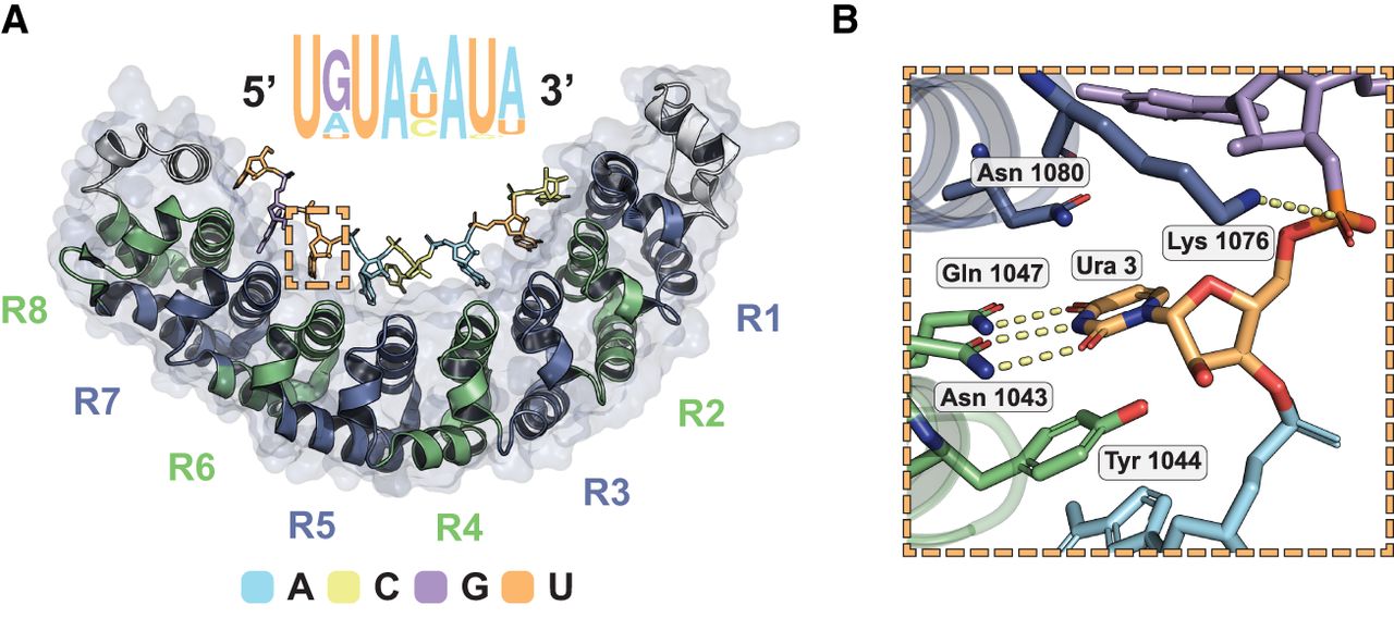
The structure of RNA bound to human Pumilio, a prototypical RNA-binding protein. (A) Crystal structure of human Pumilio 1 (PDB ID: 3Q0P) bound to a consensus RNA sequence. The Pumilio RNA-binding domain consists of eight helical PUF repeats forming a comma-shaped structure that binds RNA on its interior. Each PUF repeat (R1–R8) interacts with a single nucleobase. Binding is antiparallel, with the C terminus of Pumilio interacting with the 5′ end of the RNA sequence motif. Human Pumilio consensus motif presented, with letter size indicating in vivo sequence preference. Dashed box region enlarged. (A) Adenine, (G) guanine, (C) cytidine, (U) uridine. (B) Molecular detail of a single PUF repeat (R6) binding to a single nucleobase (uridine). A PUF repeat amino acid triplet binds to preferred bases through hydrogen bonding and stacking. Images by PyMOL.




