


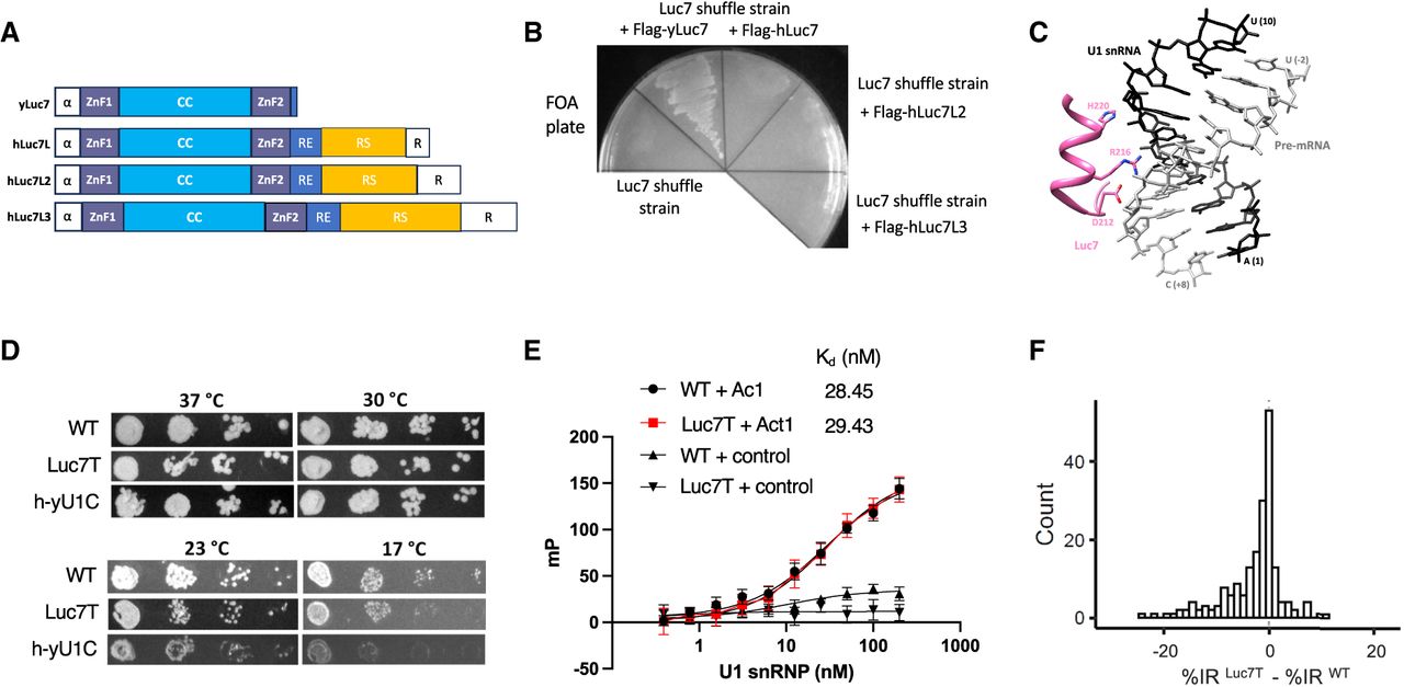
The Luc7 triple mutant has no significant effect on splicing. (A) Domain organization of the yLuc7 and hLuc7 homologs. α represents α-helix. CC represents a coiled-coil region. RE, RS, and R represent Arg/Glu, Arg/Ser, and Arg-rich regions. (B) hLuc7 homologs could not replace the yLuc7, as demonstrated by the lack of growth on a FOA plate after hLuc7Ls were introduced into the yLuc7 shuffle strain. (C) The cryo-EM structure of the yeast E complex (Li et al. 2019) indicated that residues D212, R216, and H220 were in close proximity of the U1–5′ ss RNA duplex. (D) Spot assay with 10× serial dilution showed Luc7T had similar growth as the parental WT strain, with a very mild growth retardation at 17°C that is less severe than h-yU1C. (E) FP experiments demonstrated that U1 snRNPLuc7T and U1 snRNP WT had indistinguishable binding affinity to the 5′ ss oligo. The control oligo is the 5′ end of U1 snRNA with the sequence AUACUUACCU. FP values are baseline (oligo alone) subtracted to better align and compare the two binding curves. (F) Luc7T had no significant effect on splicing. None of the IR differences observed between the Luc7T and WT was significantly larger (FRD < 0.05) than the IR differences observed among the three biological replicates for WT or Luc7T.




