


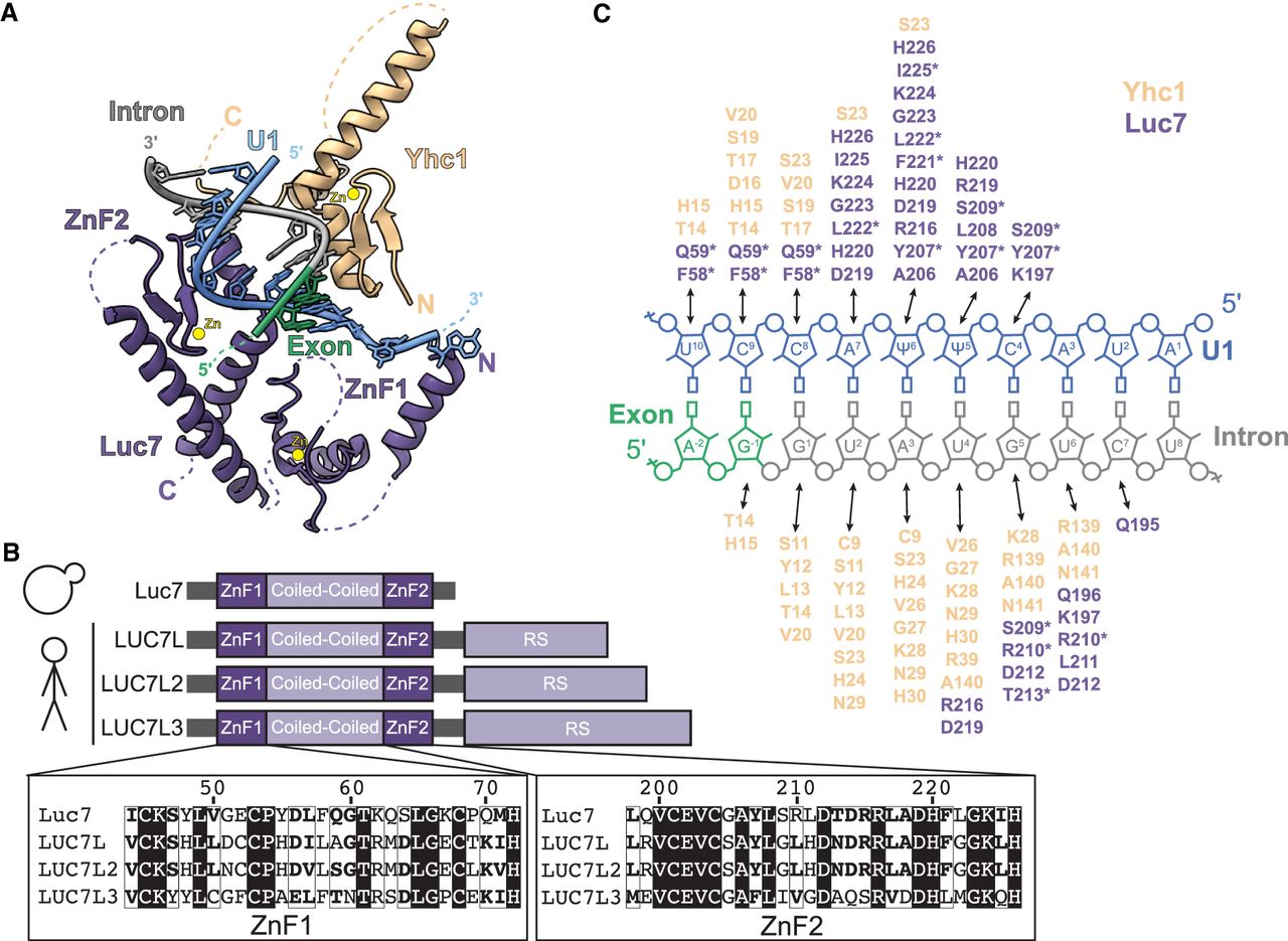
FIGURE 1.
The U1:5′ss duplex contacts Yhc1/U1-C and Luc7/LUC7L. (A) U1:5′ss base-pairing is stabilized by the Yhc1 and Luc7 ZnF domains in the structure of the yeast prespliceosome (PDB 6G90). Luc7 ZnF2 contacts the 5′ss duplex, whereas ZnF1 lies in the direction of exon 1, which are both partially unresolved. (B) Schematic showing LUC7 and the three human paralogs. A sequence alignment shows the similarity of the ZnF domains. (C) Schematic showing Yhc1 (beige) and Luc7 (purple) residues located within 6 Å of the U1:5′ss duplex (blue/gray). Asterisks indicate residues that are not conserved between yeast and human Luc7 proteins.




