


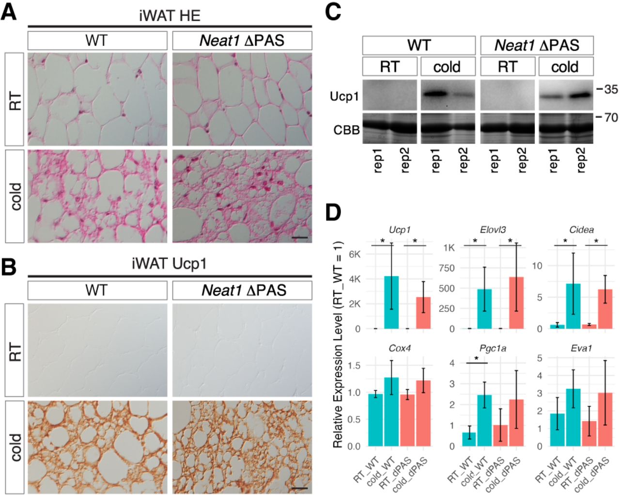
Beige adipocyte differentiation appears normal in Neat1 ΔPAS Mice. (A) Nomarski images of H&E-stained paraffin sections of iWAT derived from WT and Neat1 ΔPAS mice reared under normal ambient temperature (RT) and cold temperature (cold) for 1 week. (B) Immunohistochemical detection of Ucp1 in iWAT derived from WT and Neat1 ΔPAS mice reared under normal (RT) and cold temperature (cold) for 1 week. (C) Western blot analyses of Ucp1 expression in iWAT of WT and Neat1 ΔPAS animals. Two biological replicates (rep1 and rep2) were used for each condition. CBB-stained gels served as a loading control. (D) Expression of mRNAs for beige cell markers Ucp1, Elovl3, Cidea, Cox4, Pgc1a, and Eva1. The asterisks indicate statistically significant changes: (*) P < 0.05, bootstrap test, 1000 surrogates, n = 3 per group. Scale bar, 20 µm.




