


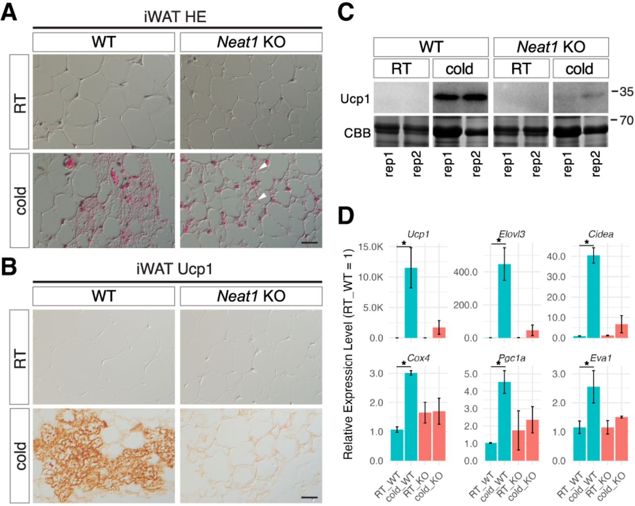
Impaired beige adipocyte differentiation in Neat1 KO mice. (A) Nomarski images of H&E-stained paraffin sections of iWAT derived from WT and Neat1 KO mice reared under normal ambient temperature (RT) and cold temperature (cold) for 1 week. Beige cells are identifiable by numerous small multilocular lipid droplets in the cytoplasm forming mesh-like structures. (B) Immunohistochemical detection of Ucp1 in iWAT derived from WT and Neat1 KO mice reared under normal (RT) and cold temperature (cold) for 1 week. Note that Ucp1 expression is strongly upregulated in WT iWAT upon cold exposure, which is absent in Neat1 KO mice. (C) Western blot analyses of Ucp1 expression in iWAT of WT and Neat1 KO animals. Two biological replicates (rep1 and rep2) were used for each condition. Coomassie brilliant blue (CBB)-stained gels served as a loading control. (D) Expression of mRNAs for beige cell markers Ucp1, Elovl3, Cidea, Cox4, Pgc1a, and Eva1. Note that these markers were upregulated upon cold exposure in WT mice, which is attenuated in Neat1 KO mice. The asterisks indicate statistically significant changes: (*) P < 0.05, bootstrap test, 1000 surrogates, n = 2 for RT and n = 3 for cold. Scale bar, 20 µm.




