


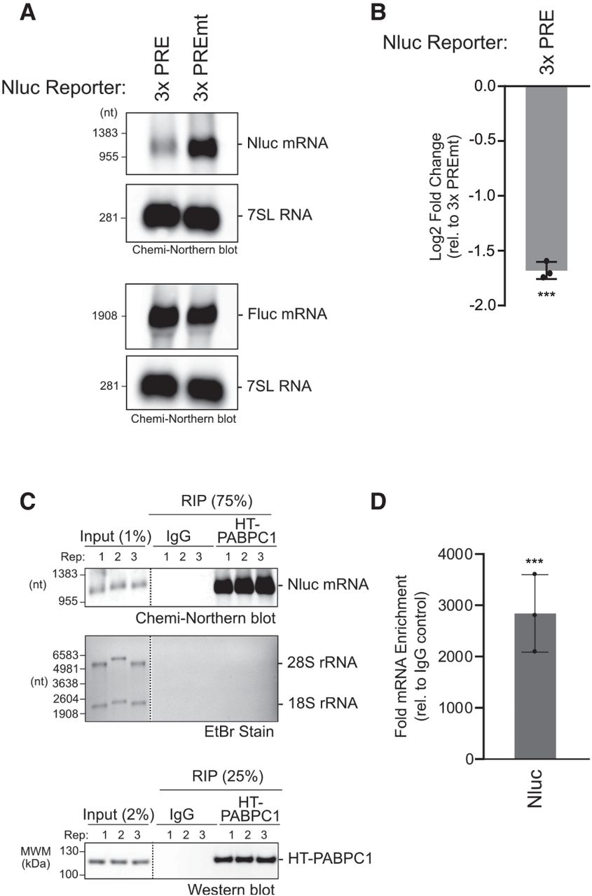
Chemi-Northern reproducibly measures mRNA regulation and binding by RNA-binding proteins. (A) Regulation of the Nluc reporter mRNA with three wild-type Pumilio response elements (3×PRE) in its 3′UTR was compared to the mutant version (3×PREmt) using Chemi-Northern. The PREs are specifically recognized by endogenous sequence-specific RNA-binding proteins, PUM1 and PUM2, which repress by causing degradation of the mRNA, as previously documented (Van Etten et al. 2012; Bohn et al. 2018; Wolfe et al. 2020; Enwerem et al. 2021). Reporters were expressed in HCT116 cells. The Fluc mRNA served as a control for transfection efficiency (the Fluc probe has 7% biotinylated nucleotides). Nluc mRNA was detected at 5 sec exposure and Fluc mRNA at 10 sec. The 7SL RNA served as an internal control for equivalent loading of each gel and was imaged at 5 sec exposure time. (B) Quantitation of Chemi-Northern in panel A and three additional biological replicates. Log2 fold change of the Nluc 3×PRE reporter was determined relative to the mutant version, Nluc 3×PREmt, in each sample and was calculated as described in Materials and Methods. Mean and SD values are plotted, along with the values of each of the replicates. Statistical significance (***) P = 0.0005 based on unpaired two-tailed t-test. (C) Nluc mRNA is enriched by RIP of human RNA-binding protein PABPC1, expressed as a fusion to Halotag (HT-PABPC1) from HCT116 cells, wherein the endogenous PABPC1 and PABPC4 paralogs were depleted (see Materials and Methods). The top image shows the Chemi-Northern detection of the polyadenylated Nluc mRNA at 40 sec exposure in the input samples and the robust enrichment in the HT-PABPC1 RIP samples, but not the IgG negative control RIPs. The middle image shows the EtBr-stained denaturing formaldehyde–MOPS agarose gel from input and RIP samples. Ribosomal RNAs are clearly visible in the inputs, but not in RIP samples. The image at the bottom shows the western blot of the HT-PABPC1 protein, demonstrating its presence in the input samples and enrichment in the RIP samples. Dashed vertical lines in the panels indicate that the images were cropped to show relevant lanes. For each image, all of the lanes are from the same blot and exposure. (D) Quantitation of the Chemi-Northern in C demonstrates significant, reproducible detection of Nluc mRNA in the PABPC1 RIP samples. Nluc RNA signal intensity in each RIP sample was normalized to its corresponding input sample, then the fold enrichment in the PABPC1 RIP was calculated relative to the IgG negative control. The mean and SD values are plotted, along with the values of each of the three biological replicates. Statistical significance (***) P = 0.0005 based on an unpaired two-tailed t-test.




