


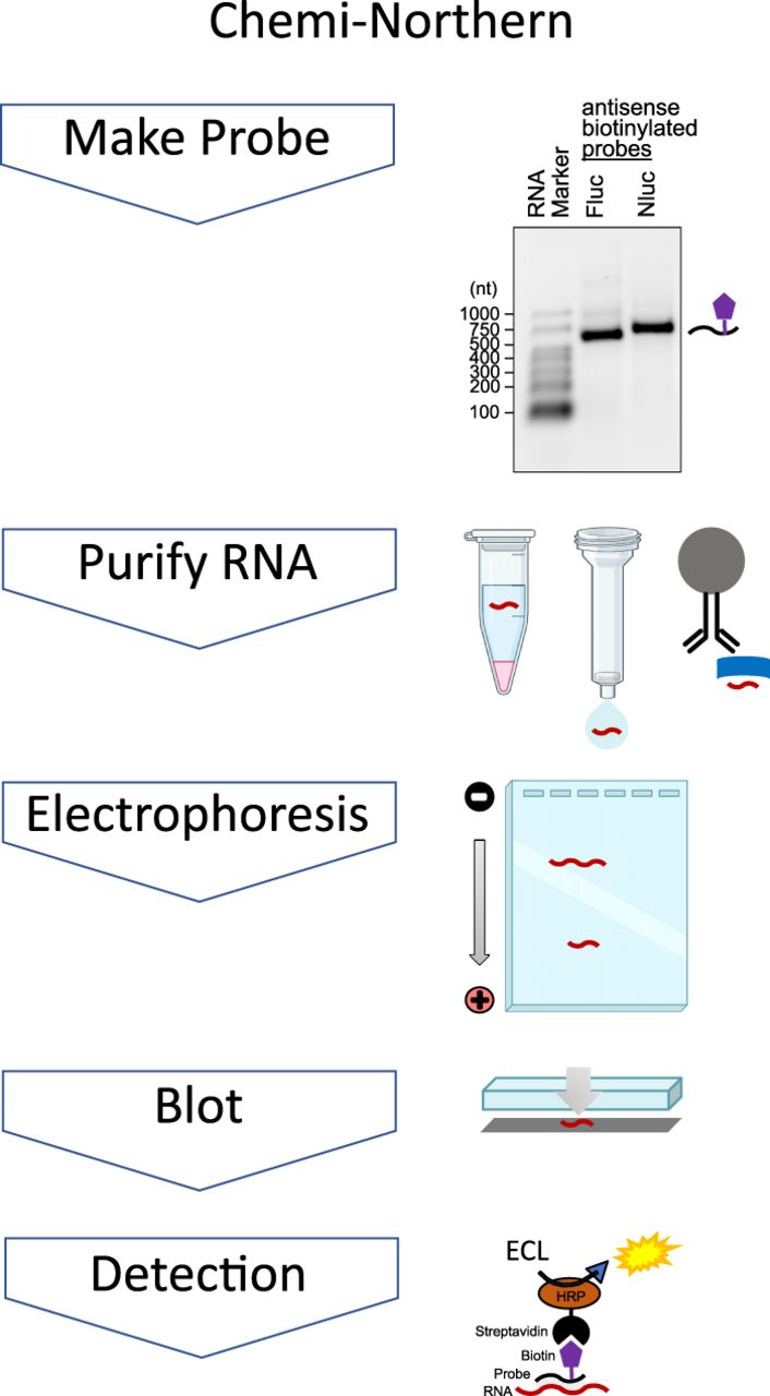
Overview of RNA detection by Chemi-Northern method. The Chemi-Northern protocol consists of five steps: (1) Generation of biotinylated (purple pentagon) antisense nucleic acid probe (black line). As an example, an ethidium bromide (EtBr)-stained denaturing formaldehyde–MOPS agarose gel of the firefly luciferase (Fluc) and nanoluciferase (Nluc) RNA probes used in this study are shown on the right. An RNA size marker is included on the left, with sizes indicated in nucleotides (nt). (2) Extraction and purification of RNA (red line). As shown on the right, RNAs can be purified from diverse sources using a variety of methods including organic phase separation (e.g., TRIzol reagent), spin column, or bead-based methods (e.g., RNA coimmunoprecipitation [RIP] assays). (3) Separation of RNA by charge-to-length ratio by denaturing formaldehyde–MOPS agarose gel electrophoresis. (4) Blotting transfer and immobilization of RNA to positively charged nylon membrane. (5) Visualization of target RNA hybridized to the biotinylated probe using streptavidin–HRP conjugate and enhanced chemiluminescence (ECL) detection. Gel, column, and test tube icons were created by Biorender.com under academic license agreement NE25MGJNLD.




