


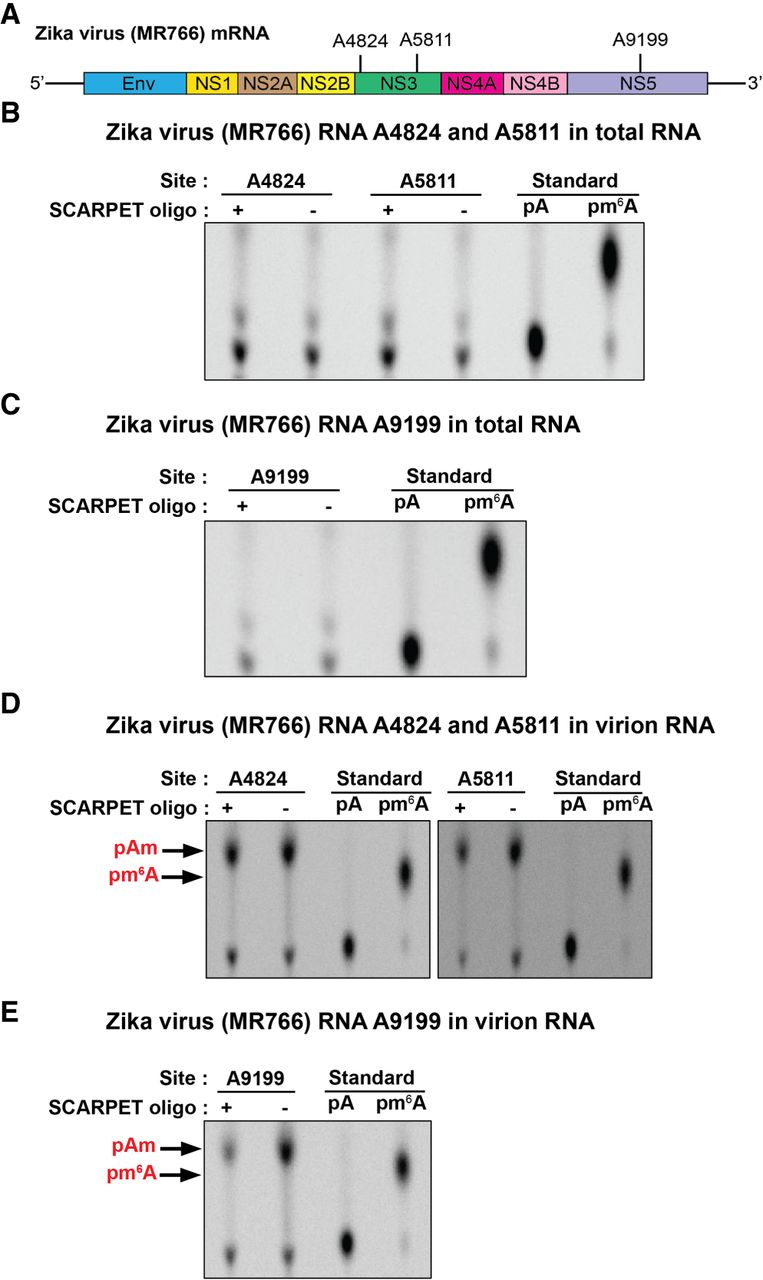
m6A not detectable at selected sites in Zika RNA. (A) Structure of Zika virus (MR766) mRNA depicting A4824, A5811, and A9199 sites. All these sites are in common GGACU consensus site for m6A and are located within m6A peaks that were previously detected using MeRIP-seq m6A mapping (Lichinchi et al. 2016; Gokhale et al. 2020). (B,C) SCARPET results for A4824, A5811, and A9199 sites using DNase I-treated and rRNA-depleted RNA isolated from Huh-7.5 hepatocellular adenocarcinoma cells infected with MR766 Zika virus 72 h postinfection. SCARPET showed a lack of detectable m6A at all three sites. (D,E) SCARPET results for A4824, A5811, and A9199 sites using DNase I-treated and rRNA-depleted RNA isolated from virion particles collected from Zika virus MR766-infected Vero E6 (African green monkey kidney) cells 72 h postinfection. SCARPET showed a prominent Am spot that was independent of the SCARPET oligonucleotide but no m6A spot. The Am spot likely reflects low but measurable ligation of the hairpin oligonucleotide to the 5′ end of the Zika genomic RNA which begins with Am (Coutard et al. 2017). 32P-labeled m6A and A nucleosides were run as TLC standards in B–D.




