


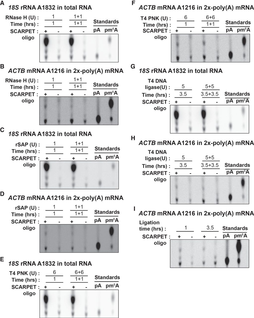
Optimization of SCARPET assay steps. (A,B) RNase H step (A) and (B) optimization of the RNase H-assisted cleavage step in SCARPET by either doubling the amount of RNase H enzyme or the time of incubation compared to standard RNase H reaction conditions (1 U RNase H, 1 h incubation at 44°C). Neither increasing the amount of enzyme nor time of incubation increased total levels of A and m6A at either of these sites. (C,D) rSAP step (C) and (D) Optimization of dephosphorylation step by rSAP enzyme in SCARPET by either doubling the amount of rSAP enzyme or the time of incubation. Compared to standard rSAP reaction conditions (1 U rSAP, 1 h incubation at 44°C), neither increasing the amount of enzyme nor time of incubation resulted in increased levels of A and m6A at either of these sites. (E,F) PNK radiolabeling step (E) and (F) Optimization of radiolabeling by T4 PNK enzyme in SCARPET by either doubling the amount of T4 PNK enzyme or the time of incubation. Compared to standard radiolabeling reaction conditions (6 U T4 PNK, 10 µCi [γ-32P]-ATP, 1 h incubation at 37°C), neither increasing the amount of enzyme nor time of incubation resulted in increased levels of A and m6A at either of these sites. (G,H) Self-splinting hairpin-assisted ligation step (G) and (H) Optimization of ligation step by T4 DNA ligase enzyme in SCARPET by either doubling the amount of T4 DNA ligase enzyme or the time of incubation. Compared to standard ligation reaction conditions (5 U T4 DNA ligase, 3.5 h incubation at 37°C), neither increasing the amount of enzyme nor time of incubation resulted in increased levels of A and m6A at either of these sites. Notably, prolonged ligation seemed to increase the amount of the A spot, which likely reflects increased nonspecific background ligation. (I) Shortening the ligation time to only 1 h leads to a decrease in the background level of A suggesting that 1 h was sufficient for ligation, and prolonged ligation times could increase background signals. The RNA input for all these experiments was 1 µg total RNA for 18S rRNA A1832 site and 1 µg 2×-poly(A) mRNA for ACTB mRNA A1216 site from HEK293T cells and 20 ng of SCARPET oligo was used in all experiments except in the controls where it was omitted. 32P-labeled m6A and A nucleosides were run as TLC standards in A–I.




